Department of Human Biology, NUTRIM School of Nutrition and Translational Research in Metabolism, Maastricht University Medical Centre+, Maastricht, The Netherlands.
Rehabilitation Research Center, BIOMED Biomedical Research Institute, Faculty of Medicine and Life Sciences, Hasselt University, Diepenbeek, Belgium.
Sci Rep. 2018 Mar 16;8(1):4677. doi: 10.1038/s41598-018-22962-x.
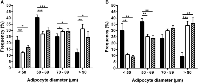
Obesity is associated with a disturbed adipose tissue (AT) function characterized by adipocyte hypertrophy, an impaired lipolysis and pro-inflammatory phenotype, which contributes to insulin resistance (IR). We investigated whether AT phenotype in different AT depots of obese individuals with and without type 2 diabetes mellitus (T2DM) is associated with whole-body IR. Subcutaneous (SC) and visceral (V) AT biopsies from 18 lean, 17 obese and 8 obese T2DM men were collected. AT phenotype was characterized by ex vivo measurement of basal and stimulated lipolysis (mature adipocytes), adipocyte size distribution (AT tissue sections) and AT immune cells (flow cytometry). In VAT, mean adipocyte size, CD45 leukocytes and M1 macrophages were significantly increased in both obese groups compared to lean individuals. In SCAT, despite adipocyte hypertrophy, no significant differences in immune cell populations between groups were found. In SCAT, multiple linear regression analysis showed that none of the AT phenotype markers independently contributed to HOMA-IR while in VAT, mean adipocyte size was significantly related to HOMA-IR. In conclusion, beside adipocyte hypertrophy in VAT, M1 macrophage- or B-cell-mediated inflammation, may contribute to IR, while inflammation in hypertrophic SCAT does not seem to play a major role in IR.
肥胖与脂肪组织(AT)功能障碍有关,其特征为脂肪细胞肥大、脂解受损和促炎表型,这导致了胰岛素抵抗(IR)。我们研究了肥胖个体中不同 AT 部位的 AT 表型是否与全身 IR 有关,这些肥胖个体中有些患有 2 型糖尿病(T2DM),有些没有。从 18 名瘦人、17 名肥胖者和 8 名肥胖 T2DM 男性中采集了皮下(SC)和内脏(V)AT 活检。通过对基础和刺激脂解(成熟脂肪细胞)、脂肪细胞大小分布(AT 组织切片)和 AT 免疫细胞(流式细胞术)的离体测量来表征 AT 表型。在 VAT 中,与瘦人相比,两组肥胖者的平均脂肪细胞大小、CD45 白细胞和 M1 巨噬细胞均显著增加。在 SCAT 中,尽管存在脂肪细胞肥大,但各组之间的免疫细胞群没有明显差异。在 SCAT 中,多元线性回归分析显示,没有任何 AT 表型标志物可独立贡献 HOMA-IR,而在 VAT 中,平均脂肪细胞大小与 HOMA-IR 显著相关。总之,除了 VAT 中的脂肪细胞肥大外,M1 巨噬细胞或 B 细胞介导的炎症可能导致 IR,而肥大的 SCAT 中的炎症似乎在 IR 中不起主要作用。