1 Department of Anesthesiology, University of Texas Medical Branch , Galveston, Texas.
2 Department of Neuroscience and Cell Biology, University of Texas Medical Branch , Galveston, Texas.
J Neurotrauma. 2018 Jul 1;35(13):1510-1522. doi: 10.1089/neu.2017.5249. Epub 2018 Apr 30.
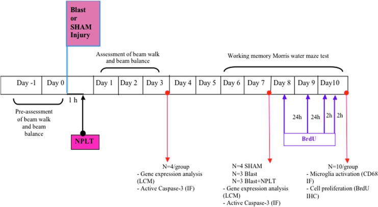
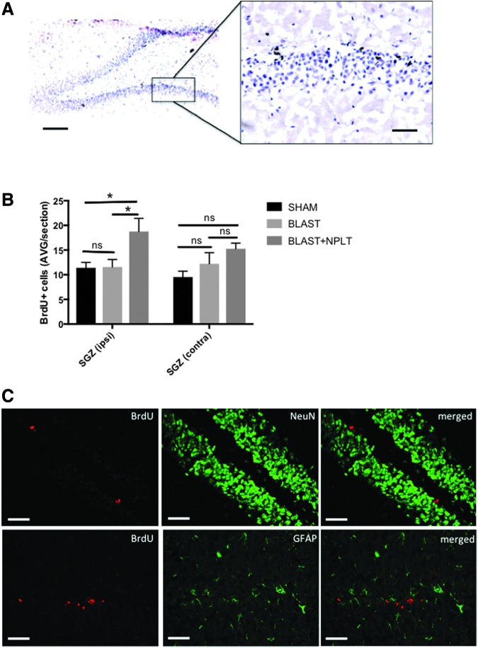
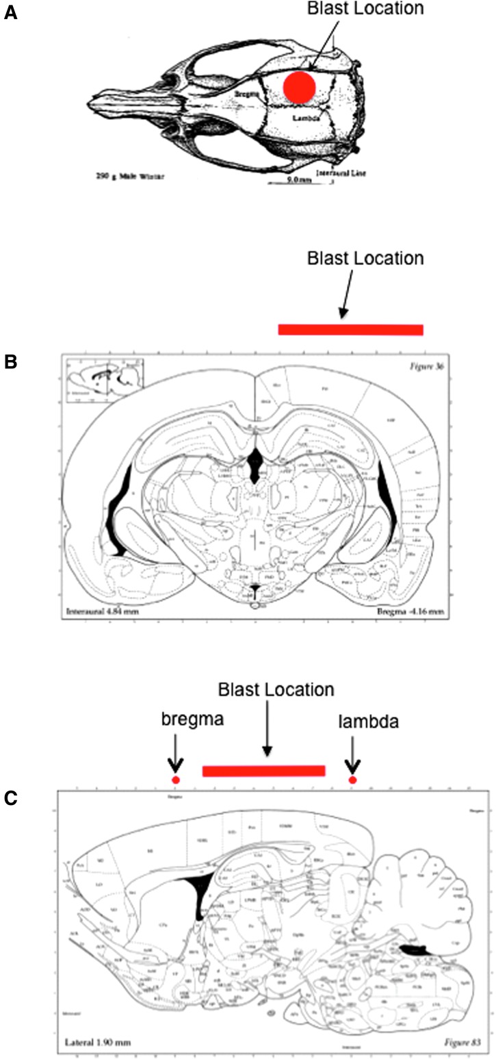
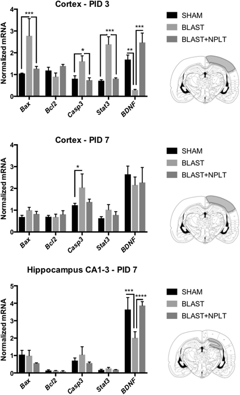
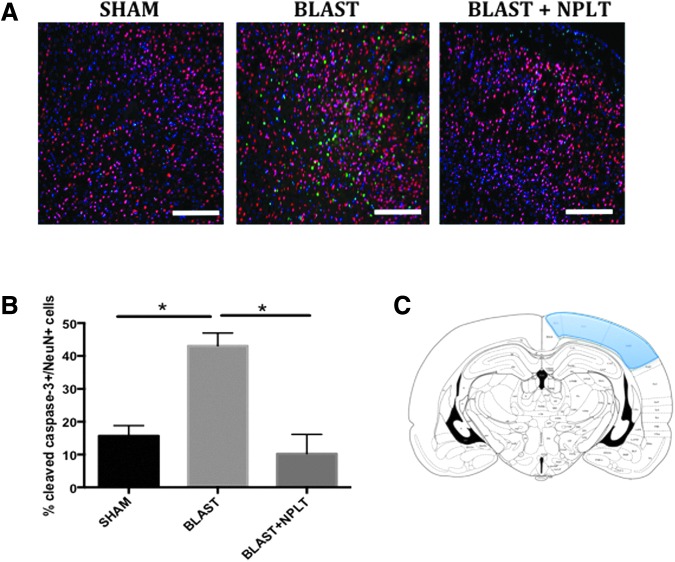
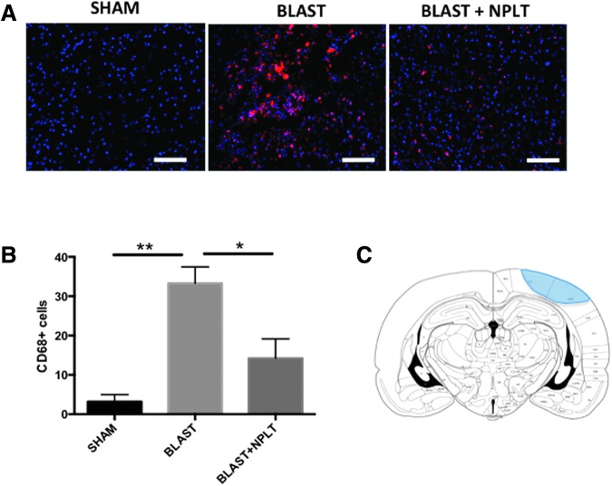
We have developed a novel, non-invasive nano-pulsed laser therapy (NPLT) system that combines the benefits of near-infrared laser light (808 nm) and ultrasound (optoacoustic) waves, which are generated with each short laser pulse within the tissue. We tested NPLT in a rat model of blast-induced neurotrauma (BINT) to determine whether transcranial application of NPLT provides neuroprotective effects. The laser pulses were applied on the intact rat head 1 h after injury using a specially developed fiber-optic system. Vestibulomotor function was assessed on post-injury days (PIDs) 1-3 on the beam balance and beam walking tasks. Cognitive function was assessed on PIDs 6-10 using a working memory Morris water maze (MWM) test. BDNF and caspase-3 messenger RNA (mRNA) expression was measured by quantitative real-time PCR (qRT-PCR) in laser-captured cortical neurons. Microglia activation and neuronal injury were assessed in brain sections by immunofluorescence using specific antibodies against CD68 and active caspase-3, respectively. In the vestibulomotor and cognitive (MWM) tests, NPLT-treated animals performed significantly better than the untreated blast group and similarly to sham animals. NPLT upregulated mRNA encoding BDNF and downregulated the pro-apoptotic protein caspase-3 in cortical neurons. Immunofluorescence demonstrated that NPLT inhibited microglia activation and reduced the number of cortical neurons expressing activated caspase-3. NPLT also increased expression of BDNF in the hippocampus and the number of proliferating progenitor cells in the dentate gyrus. Our data demonstrate a neuroprotective effect of NPLT and prompt further studies aimed to develop NPLT as a therapeutic intervention after traumatic brain injury (TBI).
我们开发了一种新型的非侵入性纳米脉冲激光治疗(NPLT)系统,该系统结合了近红外激光(808nm)和超声(光声)波的优点,这些波是通过组织内的每个短激光脉冲产生的。我们在爆炸诱导的神经创伤(BINT)大鼠模型中测试了 NPLT,以确定经颅应用 NPLT 是否提供神经保护作用。激光脉冲通过专门开发的光纤系统在损伤后 1 小时应用于完整的大鼠头部。前庭运动功能在损伤后 1-3 天(PID)在梁平衡和梁行走任务上进行评估。使用工作记忆 Morris 水迷宫(MWM)测试在 PID 6-10 上评估认知功能。通过定量实时 PCR(qRT-PCR)测量激光捕获的皮质神经元中的 BDNF 和半胱天冬酶-3 信使 RNA(mRNA)表达。使用针对 CD68 和活性半胱天冬酶-3 的特异性抗体通过免疫荧光在脑切片中评估小胶质细胞激活和神经元损伤。在前庭运动和认知(MWM)测试中,NPLT 治疗的动物表现明显优于未治疗的爆炸组,与假手术动物相似。NPLT 上调编码 BDNF 的 mRNA,并下调皮质神经元中的促凋亡蛋白半胱天冬酶-3。免疫荧光表明,NPLT 抑制小胶质细胞激活并减少表达激活的半胱天冬酶-3 的皮质神经元数量。NPLT 还增加了海马中的 BDNF 表达和齿状回中增殖祖细胞的数量。我们的数据表明 NPLT 具有神经保护作用,并促使进一步研究旨在开发 NPLT 作为创伤性脑损伤(TBI)后的治疗干预措施。