Department of Rehabilitation, the First Affiliated Hospital of Chongqing Medical University, Chongqing, China.
Department of Rehabilitation, Southwest University Hospital, Chongqing, China.
Cancer Med. 2018 May;7(5):1908-1920. doi: 10.1002/cam4.1418. Epub 2018 Mar 25.
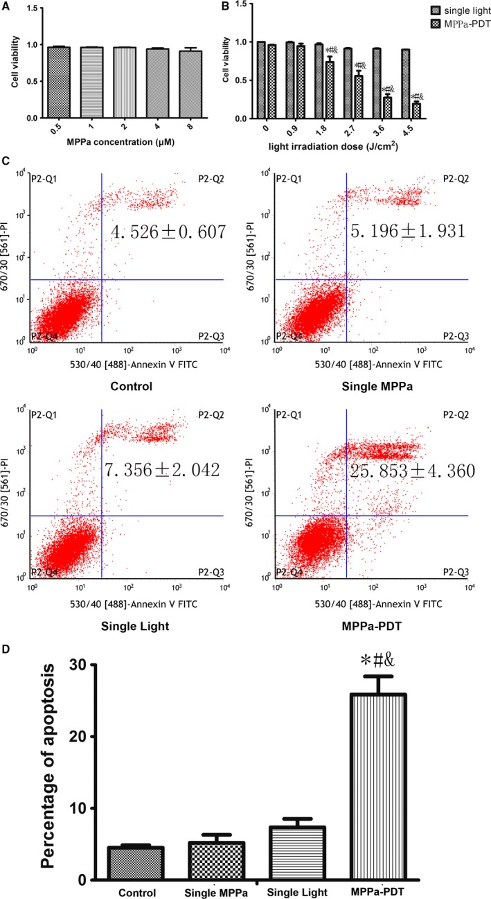
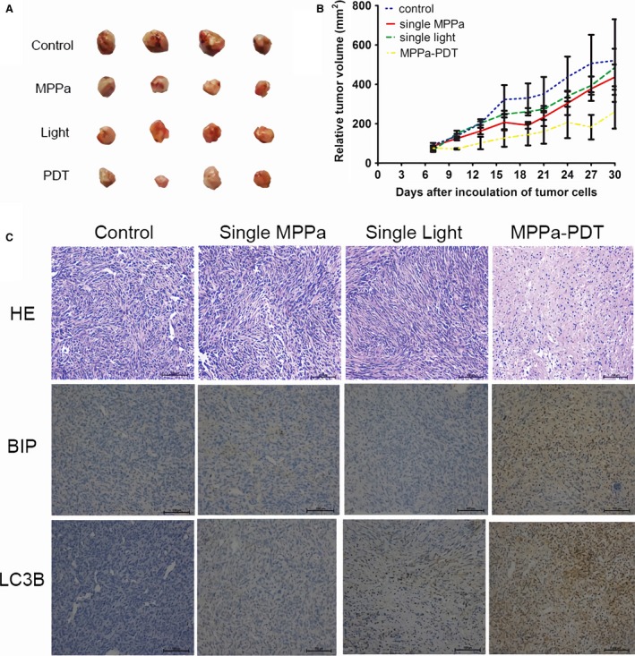
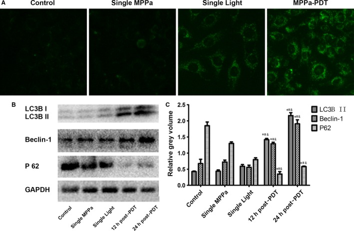
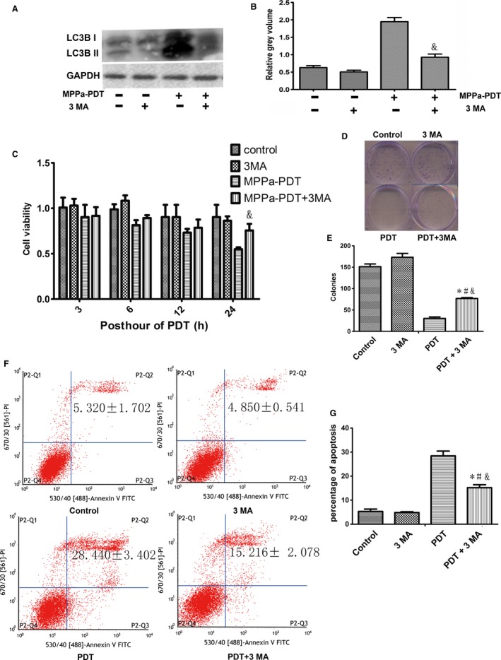
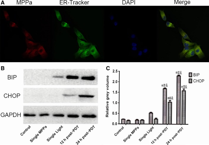
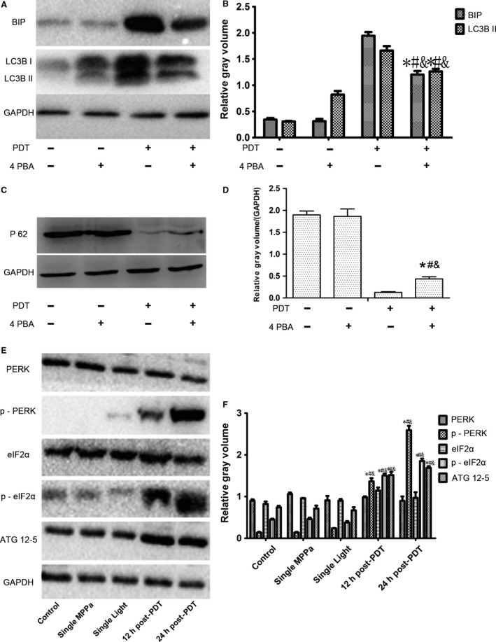
Autophagy and ER stress participated in the inhibition of MPPa-PDT on tumor growth, but the molecular links between them remain undefined. We just explore the molecular mechanism between them in vitro and vivo. CCK-8 assay and flow cytometer were used to detect the cytotoxicity and mode of cell death after MPPa-PDT. Furthermore, the role of autophagy was verified in MPPa-PDT. Confocal microscopy was used to show the intracellular distribution of MPPa. ER stress markers and PERK signaling pathway were detected by western blot. While in vivo, tumor histology and immunohistochemistry were performed to show the effect of MPPa-PDT in mice. After MPPa-PDT, cells viability decreased in dose-dependent manner. Besides, the cell apoptosis increased along with the increasing of Beclin-1and LC3B II but declining of P62. When pretreated with 3-MA, LC3B II formation and the cytotoxicity declined. MPPa-PDT caused increasing of ER stress markers (GRP78, CHOP) as MPPa accumulated in ER. However, pretreatment with ER stress inhibitor 4PBA, the expression of GRP78 and LC3B II was blocked but the PERK signaling pathway activated and the expression of P62 increased. In vivo, the tumor growth was significantly inhibited by MPPa-PDT. Besides, the appearance of ER stress and autophagy was further demonstrated by immunohistochemistry. Our findings demonstrate that autophagy mediated by MPPa-PDT was regulated by ER stress, via PERK signaling pathway, to kill MDA-MB-231 cells in vitro and vivo.
自噬和 ER 应激参与了 MPPa-PDT 对肿瘤生长的抑制,但它们之间的分子联系尚不清楚。我们只是在体外和体内探索它们之间的分子机制。CCK-8 检测和流式细胞术用于检测 MPPa-PDT 后的细胞毒性和细胞死亡方式。此外,还验证了自噬在 MPPa-PDT 中的作用。共聚焦显微镜用于显示 MPPa 的细胞内分布。Western blot 用于检测 ER 应激标记物和 PERK 信号通路。在体内,通过肿瘤组织学和免疫组织化学来显示 MPPa-PDT 在小鼠中的作用。MPPa-PDT 后,细胞活力呈剂量依赖性下降。此外,随着 Beclin-1 和 LC3B II 的增加和 P62 的减少,细胞凋亡增加。当用 3-MA 预处理时,LC3B II 的形成和细胞毒性下降。当 MPPa 在 ER 中积累时,MPPa-PDT 导致 ER 应激标记物(GRP78、CHOP)的增加。然而,用 ER 应激抑制剂 4PBA 预处理时,GRP78 和 LC3B II 的表达被阻断,但 PERK 信号通路被激活,P62 的表达增加。在体内,MPPa-PDT 显著抑制了肿瘤生长。此外,免疫组织化学进一步证明了 ER 应激和自噬的出现。我们的研究结果表明,MPPa-PDT 介导的自噬通过 PERK 信号通路被 ER 应激调节,以杀死体外和体内的 MDA-MB-231 细胞。