Lu Yu-Jen, Lin Pin-Yi, Huang Pei-Han, Kuo Chang-Yi, Shalumon K T, Chen Mao-Yu, Chen Jyh-Ping
Department of Neurosurgery, Chang Gung Memorial Hospital Linkuo Medical Center and College of Medicine, Chang Gung University, Taoyuan 33305, Taiwan.
Department of Chemical and Materials Engineering, Chang Gung University, Taoyuan 33302, Taiwan.
Nanomaterials (Basel). 2018 Mar 27;8(4):193. doi: 10.3390/nano8040193.
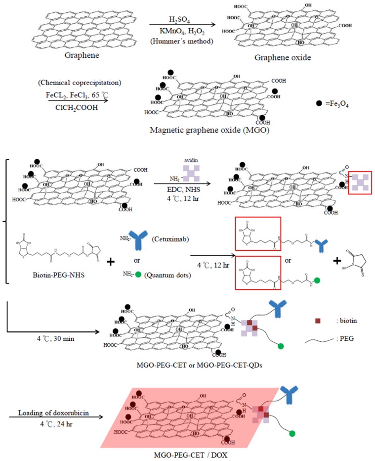
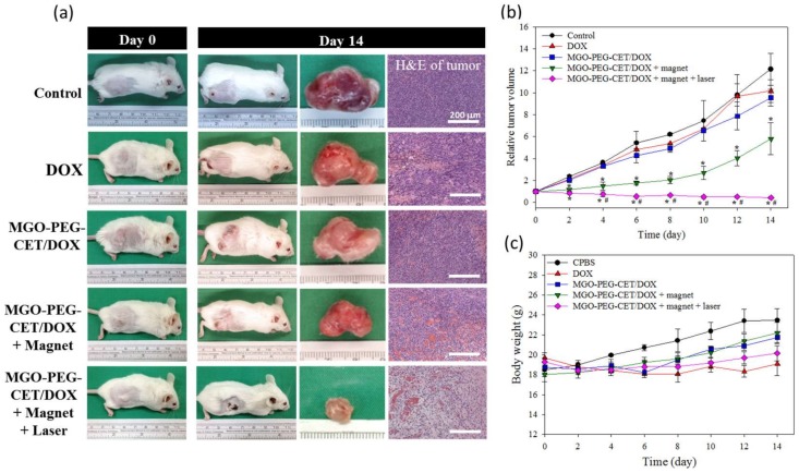
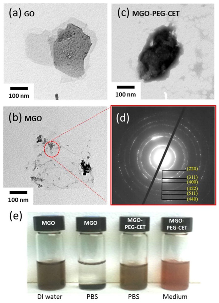
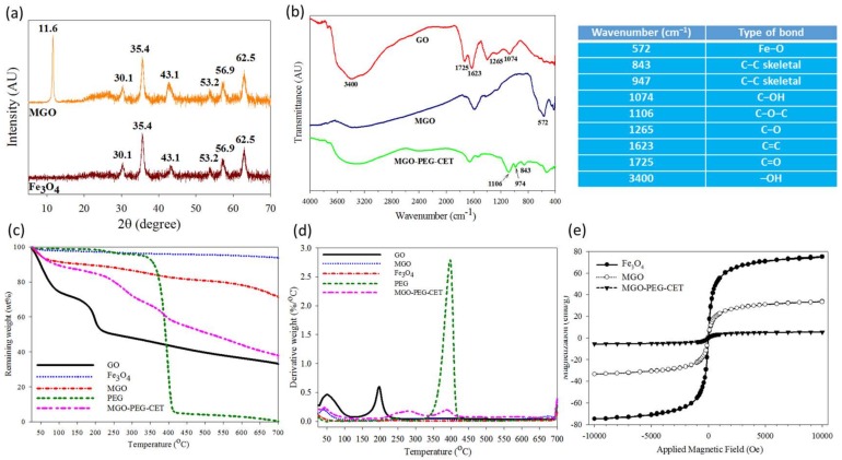
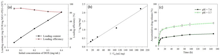
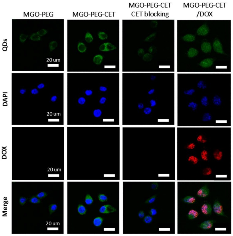
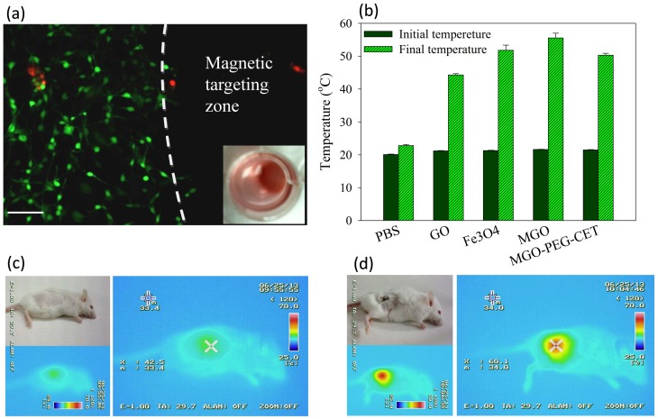
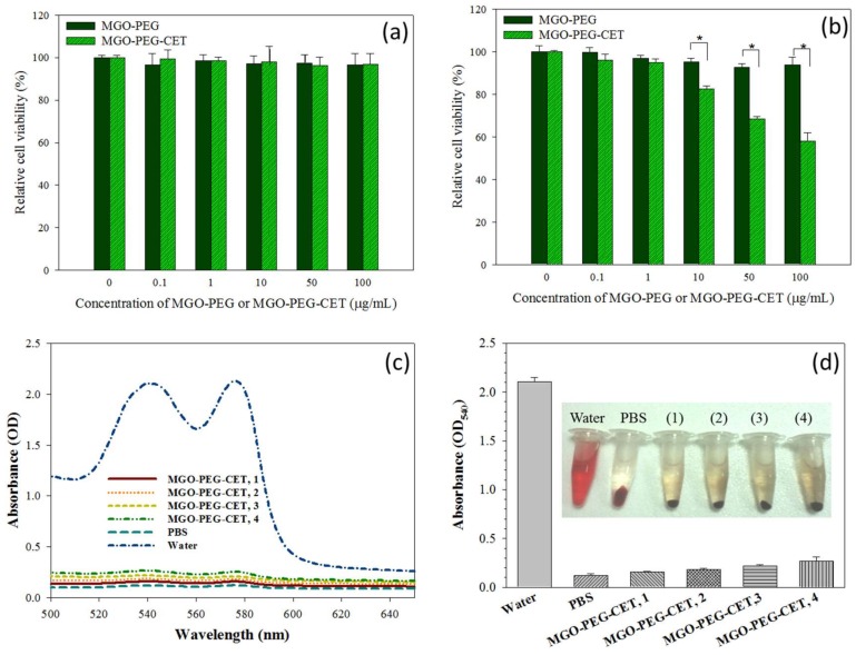
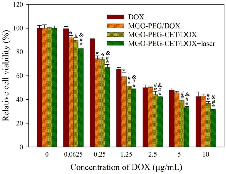
To develop a pH-sensitive dual targeting magnetic nanocarrier for chemo-phototherapy in cancer treatment, we prepared magnetic graphene oxide (MGO) by depositing Fe₃O₄ magnetic nanoparticles on graphene oxide (GO) through chemical co-precipitation. MGO was modified with polyethylene glycol (PEG) and cetuximab (CET, an epidermal growth factor receptor (EGFR) monoclonal antibody) to obtain MGO-PEG-CET. Since EGFR was highly expressed on the tumor cell surface, MGO-PEG-CET was used for dual targeted delivery an anticancer drug doxorubicin (DOX). The physico-chemical properties of MGO-PEG-CET were fully characterized by dynamic light scattering, transmission electron microscopy, X-ray diffraction, Fourier transform Infrared spectroscopy, thermogravimetric analysis, and superconducting quantum interference device. Drug loading experiments revealed that DOX adsorption followed the Langmuir isotherm with a maximal drug loading capacity of 6.35 mg/mg, while DOX release was pH-dependent with more DOX released at pH 5.5 than pH 7.4. Using quantum-dots labeled nanocarriers and confocal microscopy, intracellular uptakes of MGO-PEG-CET by high EGFR-expressing CT-26 murine colorectal cells was confirmed to be more efficient than MGO. This cellular uptake could be inhibited by pre-incubation with CET, which confirmed the receptor-mediated endocytosis of MGO-PEG-CET. Magnetic targeted killing of CT-26 was demonstrated in vitro through magnetic guidance of MGO-PEG-CET/DOX, while the photothermal effect could be confirmed in vivo and in vitro after exposure of MGO-PEG-CET to near-infrared (NIR) laser light. In addition, the biocompatibility tests indicated MGO-PEG-CET showed no cytotoxicity toward fibroblasts and elicited minimum hemolysis. In vitro cytotoxicity tests showed the half maximal inhibitory concentration (IC50) value of MGO-PEG-CET/DOX toward CT-26 cells was 1.48 µg/mL, which was lower than that of MGO-PEG/DOX (2.64 µg/mL). The IC50 value could be further reduced to 1.17 µg/mL after combining with photothermal therapy by NIR laser light exposure. Using subcutaneously implanted CT-26 cells in BALB/c mice, in vivo anti-tumor studies indicated the relative tumor volumes at day 14 were 12.1 for control (normal saline), 10.1 for DOX, 9.5 for MGO-PEG-CET/DOX, 5.8 for MGO-PEG-CET/DOX + magnet, and 0.42 for MGO-PEG-CET/DOX + magnet + laser. Therefore, the dual targeting MGO-PEG-CET/DOX could be suggested as an effective drug delivery system for anticancer therapy, which showed a 29-fold increase in therapeutic efficacy compared with control by combining chemotherapy with photothermal therapy.
为了开发一种用于癌症化学光疗的pH敏感型双靶向磁性纳米载体,我们通过化学共沉淀法将Fe₃O₄磁性纳米颗粒沉积在氧化石墨烯(GO)上制备了磁性氧化石墨烯(MGO)。用聚乙二醇(PEG)和西妥昔单抗(CET,一种表皮生长因子受体(EGFR)单克隆抗体)对MGO进行修饰,得到MGO-PEG-CET。由于EGFR在肿瘤细胞表面高表达,MGO-PEG-CET用于双靶向递送抗癌药物阿霉素(DOX)。通过动态光散射、透射电子显微镜、X射线衍射、傅里叶变换红外光谱、热重分析和超导量子干涉装置对MGO-PEG-CET的物理化学性质进行了全面表征。载药实验表明,DOX的吸附遵循Langmuir等温线,最大载药量为6.35 mg/mg,而DOX的释放依赖于pH值,在pH 5.5时比pH 7.4时释放更多的DOX。使用量子点标记的纳米载体和共聚焦显微镜,证实高表达EGFR的CT-26小鼠结肠直肠细胞对MGO-PEG-CET的细胞内摄取比MGO更有效。这种细胞摄取可以通过与CET预孵育来抑制,这证实了MGO-PEG-CET的受体介导的内吞作用。通过MGO-PEG-CET/DOX的磁引导在体外证明了对CT-26的磁靶向杀伤,而在MGO-PEG-CET暴露于近红外(NIR)激光后,在体内和体外均可证实光热效应。此外,生物相容性测试表明MGO-PEG-CET对成纤维细胞无细胞毒性,溶血作用最小。体外细胞毒性测试表明,MGO-PEG-CET/DOX对CT-26细胞的半数最大抑制浓度(IC50)值为1.48 µg/mL,低于MGO-PEG/DOX(2.64 µg/mL)。通过近红外激光光热疗法联合使用后,IC50值可进一步降至1.17 µg/mL。在BALB/c小鼠皮下植入CT-26细胞,体内抗肿瘤研究表明,第14天的相对肿瘤体积,对照组(生理盐水)为12.1,DOX组为10.1,MGO-PEG-CET/DOX组为9.5,MGO-PEG-CET/DOX+磁铁组为5.8,MGO-PEG-CET/DOX+磁铁+激光组为0.42。因此,双靶向MGO-PEG-CET/DOX可被认为是一种有效的抗癌治疗药物递送系统,通过化疗与光热疗法相结合使其治疗效果比对照组提高了29倍。