Department of Pathology and Immunology, Division of Immunobiology, Washington University School of Medicine, BJC Institute of Health, Campus Box 8118, 660 S. Euclid Avenue, St. Louis, MO, 63110, USA.
Diabetologia. 2018 Jun;61(6):1374-1383. doi: 10.1007/s00125-018-4592-4. Epub 2018 Mar 27.
AIMS/HYPOTHESIS: We studied here the interactions between the resident macrophages of pancreatic islets with beta cells and the blood vasculature. We also examined the immunological consequences of such interactions.
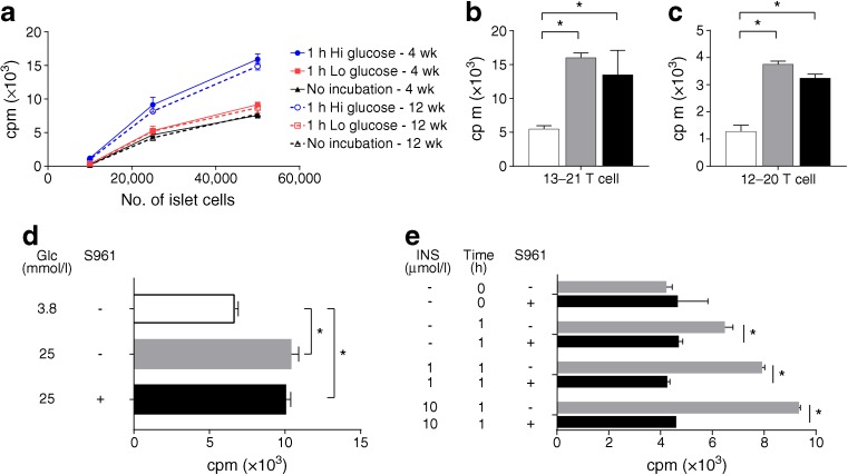
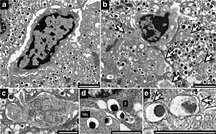
Islets were isolated from C57BL/6 mice expressing CX3C motif chemokine receptor 1-green fluorescent protein (CX3CR-GFP) and examined live by two-photon microscopy. Islets were also examined by electron microscopy to study the relationship of the intra-islet macrophages with the beta cells. In NOD.Rag1 mice and young (non-diabetic) male mice, the acquisition of beta cell granules was tested functionally by probing with CD4 T cells directed against insulin epitopes.
Two-photon microscopy showed that the islet resident macrophages were in close contact with blood vessels and had extensive filopodial activity. Some filopodia had direct access to the vessel lumen and captured microparticles. Addition of glucose at high concentration reduced the degree of filopodia sampling of islets. This finding applied to in vivo injection of glucose or to in vitro cultures. Ultrastructural examination showed the close contacts of macrophages with beta cells. Such macrophages contained intact dense core granules. Functional studies in NOD mice indicated that the macrophages presented insulin peptides to insulin-reactive T cells. Presentation was increased after glucose challenge either ex vivo or after an in vivo pulse. In agreement with the morphological findings, presentation was not affected by insulin receptor blockade.
CONCLUSIONS/INTERPRETATION: Islet resident macrophages are highly active, sampling large areas of the islets and blood contents and capturing beta cell granules. After such interactions, macrophages present immunogenic insulin to specific autoreactive T cells.
目的/假设:我们研究了胰岛固有巨噬细胞与β细胞和血管之间的相互作用。我们还检查了这种相互作用的免疫后果。
从表达 CX3C 基序趋化因子受体 1-绿色荧光蛋白 (CX3CR-GFP) 的 C57BL/6 小鼠中分离胰岛,并通过双光子显微镜实时观察。还通过电子显微镜检查胰岛,以研究胰岛内巨噬细胞与β细胞的关系。在 NOD.Rag1 小鼠和年轻(非糖尿病)雄性小鼠中,通过探测针对胰岛素表位的 CD4 T 细胞来测试β细胞颗粒的获得功能。
双光子显微镜显示胰岛固有巨噬细胞与血管密切接触,具有广泛的丝状伪足活性。一些丝状伪足直接进入血管腔并捕获微粒。高浓度葡萄糖的添加减少了胰岛丝状伪足取样的程度。这一发现适用于体内注射葡萄糖或体外培养。超微结构检查显示巨噬细胞与β细胞的紧密接触。这些巨噬细胞含有完整的致密核心颗粒。NOD 小鼠的功能研究表明,巨噬细胞将胰岛素肽呈递给胰岛素反应性 T 细胞。无论是在体外还是在体内脉冲后,葡萄糖刺激均可增加呈递。与形态学发现一致,胰岛素受体阻断不影响呈递。
结论/解释:胰岛固有巨噬细胞非常活跃,可采样胰岛和血液内容物的大片区域,并捕获β细胞颗粒。在这种相互作用之后,巨噬细胞将免疫原性胰岛素呈递给特定的自身反应性 T 细胞。