Department of Radiology, German Cancer Research Center, DKFZ, Heidelberg, Germany.
Department of Neuroradiology, University of Heidelberg Medical Center, Heidelberg, Germany.
PLoS One. 2018 Mar 29;13(3):e0193946. doi: 10.1371/journal.pone.0193946. eCollection 2018.
After the emergence of new MRI techniques such as susceptibility- and diffusion-weighted imaging (SWI and DWI) and because of specific imaging characteristics of melanoma brain metastases (MBM), it is unclear which MRI sequences are most beneficial for detection of MBM. This study was performed to investigate the sensitivity of six clinical MRI sequences in the early detection of MBM.
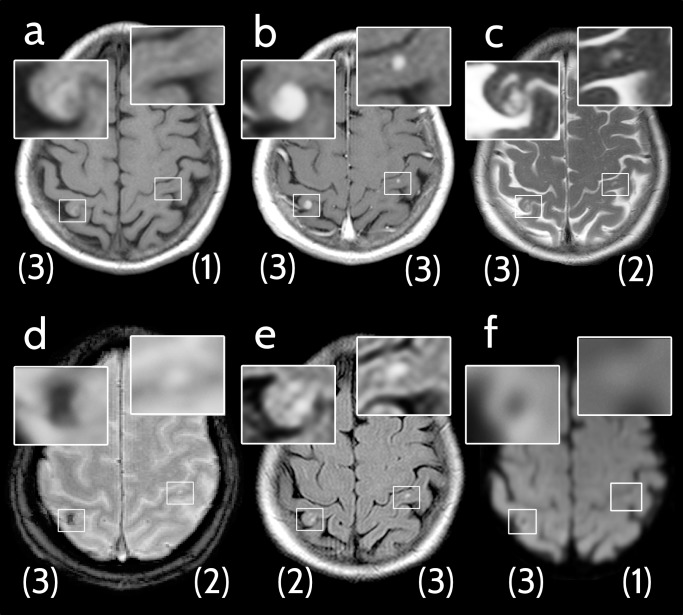
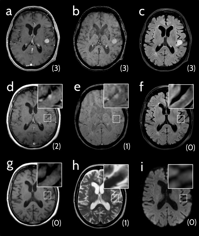
Medical records of all melanoma patients referred to our center between November 2005 and December 2016 were reviewed for presence of MBM. Analysis encompassed six MRI sequences at the time of initial diagnosis of first or new MBM, including non-enhanced T1-weighted (T1w), contrast-enhanced T1w (ceT1w), T2-weighted (T2w), T2w-FLAIR, susceptibility-weighted (SWI) and diffusion-weighted (DWI) MRI. Each lesion was rated with respect to its conspicuity (score from 0-not detectable to 3-clearly visible).
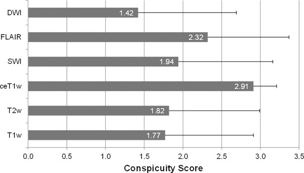
Of 1210 patients, 217 with MBM were included in the analysis and up to 5 lesions per patient were evaluated. A total of 720 metastases were assessed and all six sequences were available for 425 MBM. Sensitivity (conspicuity ≥2) was 99.7% for ceT1w, 77.0% for FLAIR, 64.7% for SWI, 61.0% for T2w, 56.7% for T1w, and 48.4% for DWI. Thirty-one (7.3%) of 425 lesions were only detectable by ceT1w but no other sequence.
Contrast-enhanced T1-weighting is more sensitive than all other sequences for detection of MBM. Disruption of the blood-brain-barrier is consistently an earlier sign in MBM than perifocal edema, signal loss on SWI or diffusion restriction.
随着新的 MRI 技术(如磁化率和弥散加权成像(SWI 和 DWI))的出现,以及由于黑色素瘤脑转移瘤(MBM)的特定成像特征,目前尚不清楚哪些 MRI 序列最有利于检测 MBM。本研究旨在探讨六种临床 MRI 序列在早期检测 MBM 中的敏感性。
回顾性分析 2005 年 11 月至 2016 年 12 月期间我院所有转诊的黑色素瘤患者的病历,以确定是否存在 MBM。分析包括初诊时首次或新发 MBM 时的六种 MRI 序列,包括非增强 T1 加权(T1w)、对比增强 T1 加权(ceT1w)、T2 加权(T2w)、T2 加权液体衰减反转恢复(T2w-FLAIR)、磁化率加权(SWI)和弥散加权(DWI)MRI。每个病变的显影程度(评分为 0-不可检测至 3-清晰可见)。
在 1210 例患者中,217 例患者存在 MBM,对每位患者的最多 5 个病灶进行了评估。共评估了 720 个转移灶,425 个 MBM 可获得所有 6 个序列。ceT1w 的灵敏度(显影≥2)为 99.7%,FLAIR 为 77.0%,SWI 为 64.7%,T2w 为 61.0%,T1w 为 56.7%,DWI 为 48.4%。425 个病灶中有 31 个(7.3%)仅可通过 ceT1w 检测到,而其他序列则无法检测到。
与其他序列相比,增强 T1 加权对 MBM 的检测更敏感。与周围水肿、SWI 上的信号丢失或弥散受限相比,血脑屏障破坏始终是 MBM 的早期征象。