Leeds Institute of Cardiovascular and Metabolic Medicine, School of Medicine, University of Leeds, Leeds, United Kingdom; and.
Centre for Rheumatology, Division of Medicine, University College London, London, United Kingdom.
FASEB J. 2018 Sep;32(9):4941-4954. doi: 10.1096/fj.201701455RR. Epub 2018 Mar 30.
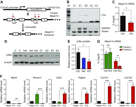
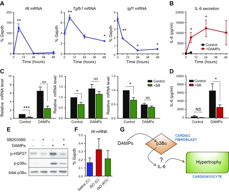
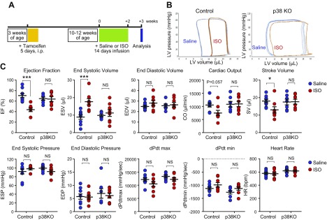
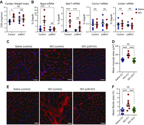
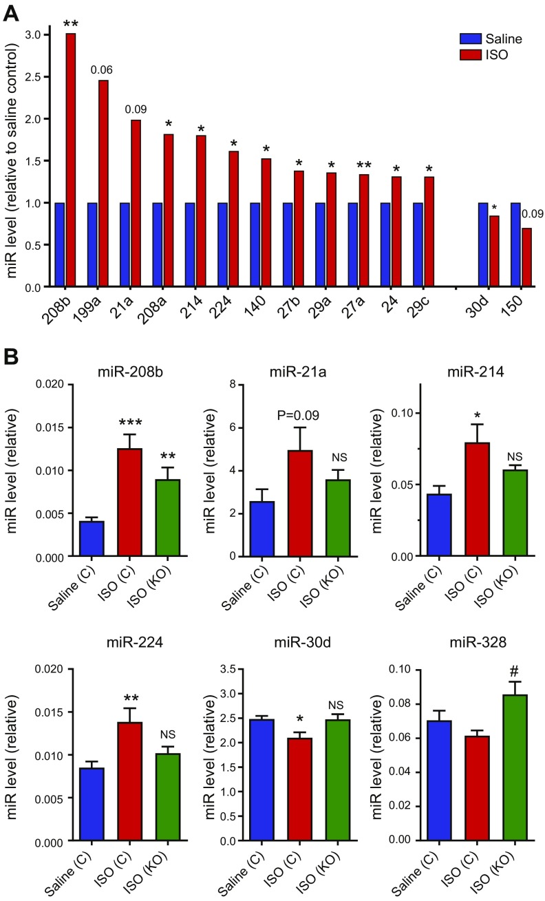
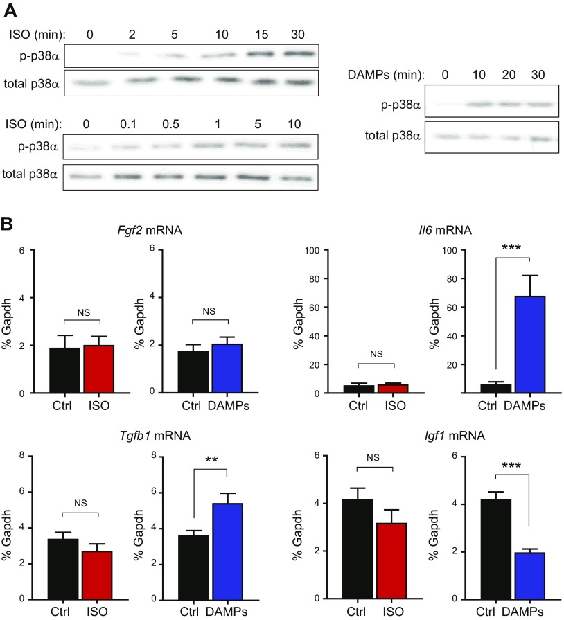
Recent studies suggest that cardiac fibroblast-specific p38α MAPK contributes to the development of cardiac hypertrophy, but the underlying mechanism is unknown. Our study used a novel fibroblast-specific, tamoxifen-inducible p38α knockout (KO) mouse line to characterize the role of fibroblast p38α in modulating cardiac hypertrophy, and we elucidated the mechanism. Myocardial injury was induced in tamoxifen-treated Cre-positive p38α KO mice or control littermates via chronic infusion of the β-adrenergic receptor agonist isoproterenol. Cardiac function was assessed by pressure-volume conductance catheter analysis and was evaluated for cardiac hypertrophy at tissue, cellular, and molecular levels. Isoproterenol infusion in control mice promoted overt cardiac hypertrophy and dysfunction (reduced ejection fraction, increased end systolic volume, increased cardiac weight index, increased cardiomyocyte area, increased fibrosis, and up-regulation of myocyte fetal genes and hypertrophy-associated microRNAs). Fibroblast-specific p38α KO mice exhibited marked protection against myocardial injury, with isoproterenol-induced alterations in cardiac function, histology, and molecular markers all being attenuated. In vitro mechanistic studies determined that cardiac fibroblasts responded to damaged myocardium by secreting several paracrine factors known to induce cardiomyocyte hypertrophy, including IL-6, whose secretion was dependent upon p38α activity. In conclusion, cardiac fibroblast p38α contributes to cardiomyocyte hypertrophy and cardiac dysfunction, potentially via a mechanism involving paracrine fibroblast-to-myocyte IL-6 signaling.-Bageghni, S. A., Hemmings, K. E., Zava, N., Denton, C. P., Porter, K. E., Ainscough, J. F. X., Drinkhill, M. J., Turner, N. A. Cardiac fibroblast-specific p38α MAP kinase promotes cardiac hypertrophy via a putative paracrine interleukin-6 signaling mechanism.
最近的研究表明,心脏成纤维细胞特异性 p38α MAPK 有助于心肌肥大的发展,但潜在的机制尚不清楚。我们的研究使用了一种新型的成纤维细胞特异性、他莫昔芬诱导的 p38α 敲除(KO)小鼠系,以表征成纤维细胞 p38α 在调节心肌肥大中的作用,并阐明了其机制。通过慢性输注β肾上腺素能受体激动剂异丙肾上腺素在他莫昔芬处理的 Cre 阳性 p38α KO 小鼠或对照同窝仔鼠中诱导心肌损伤。通过压力-容积导纳导管分析评估心脏功能,并在组织、细胞和分子水平评估心脏肥大。异丙肾上腺素输注在对照小鼠中促进了明显的心脏肥大和功能障碍(射血分数降低,舒张末期容积增加,心脏重量指数增加,心肌细胞面积增加,纤维化增加,以及胎儿基因和与肥大相关的 microRNA 上调)。成纤维细胞特异性 p38α KO 小鼠对心肌损伤表现出明显的保护作用,异丙肾上腺素诱导的心脏功能、组织学和分子标志物的改变均得到减弱。体外机制研究确定,心脏成纤维细胞通过分泌几种旁分泌因子对受损的心肌作出反应,这些因子已知可诱导心肌细胞肥大,包括 IL-6,其分泌依赖于 p38α 活性。总之,心脏成纤维细胞 p38α 有助于心肌细胞肥大和心脏功能障碍,可能通过涉及旁分泌成纤维细胞-心肌细胞 IL-6 信号的机制。