The University of Queensland Diamantina Institute, Translational Research Institute, Brisbane, QLD, Australia.
Comparative Genomics Centre, James Cook University, Townsville, QLD, Australia.
Front Immunol. 2018 Mar 16;9:483. doi: 10.3389/fimmu.2018.00483. eCollection 2018.
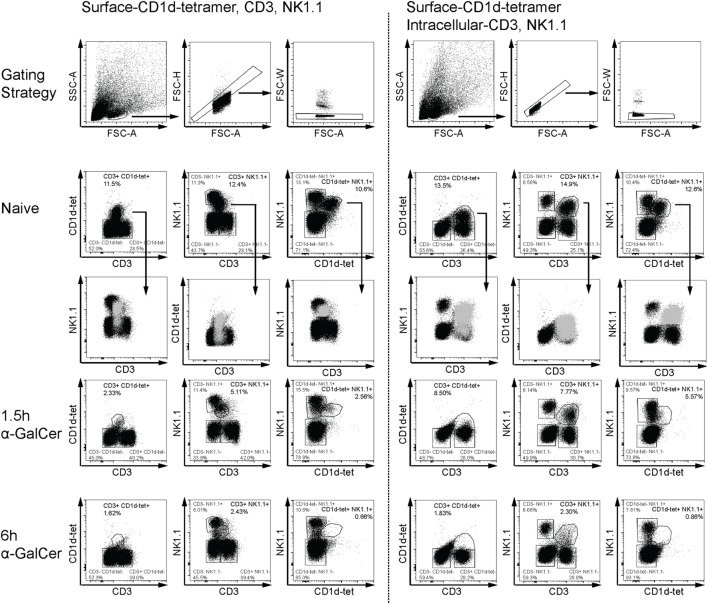
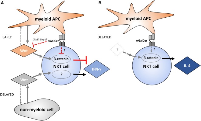
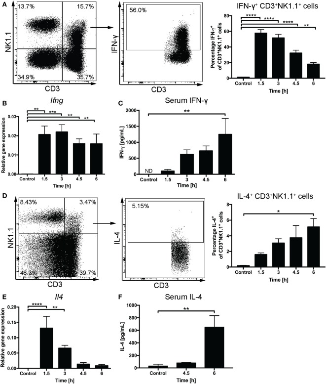
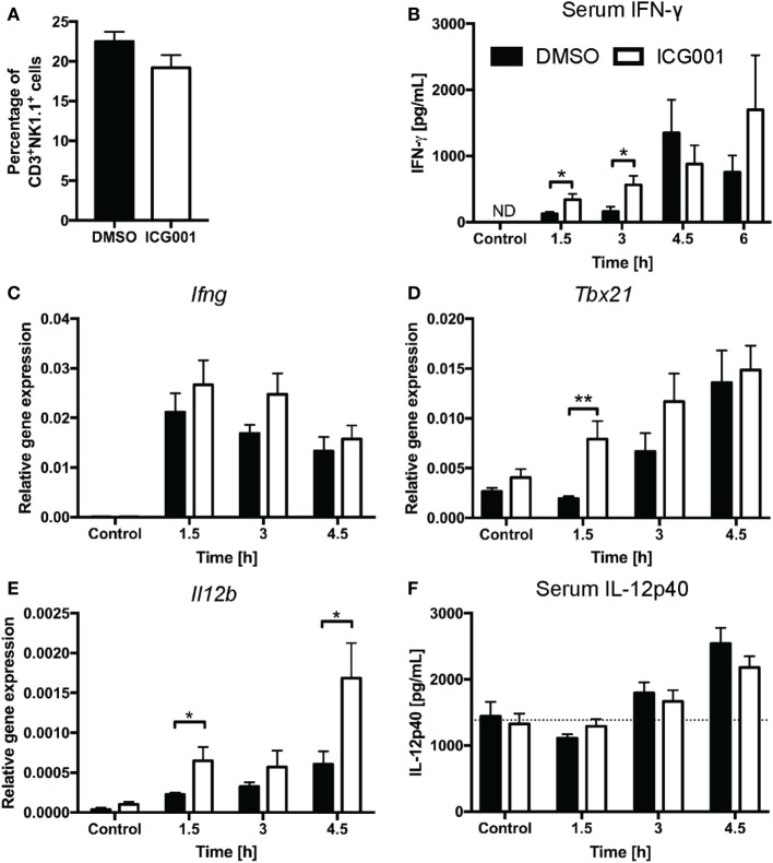
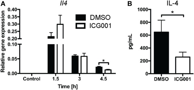
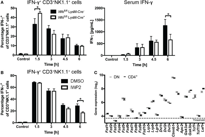
Natural killer T (NKT) cells are prominent innate-like lymphocytes in the liver with critical roles in immune responses during infection, cancer, and autoimmunity. Interferon gamma (IFN-γ) and IL-4 are key cytokines rapidly produced by NKT cells upon recognition of glycolipid antigens presented by antigen-presenting cells (APCs). It has previously been reported that the transcriptional coactivator β-catenin regulates NKT cell differentiation and functionally biases NKT cell responses toward IL-4, at the expense of IFN-γ production. β-Catenin is not only a central effector of Wnt signaling but also contributes to other signaling networks. It is currently unknown whether Wnt ligands regulate NKT cell functions. We thus investigated how Wnt ligands and β-catenin activity shape liver NKT cell functions in response to the glycolipid antigen, α-galactosylceramide (α-GalCer) using a mouse model. Pharmacologic targeting of β-catenin activity with ICG001, as well as myeloid-specific genetic ablation of , to specifically target Wnt protein release by APCs, enhanced early IFN-γ responses. By contrast, within several hours of α-GalCer challenge, myeloid-specific deficiency, as well as pharmacologic targeting of Wnt release using the small molecule inhibitor IWP-2 impaired α-GalCer-induced IFN-γ responses, independent of β-catenin activity. These data suggest that myeloid cell-derived Wnt ligands drive early Wnt/β-catenin signaling that curbs IFN-γ responses, but that, subsequently, Wnt ligands sustain IFN-γ expression independent of β-catenin activity. Our analyses in ICG001-treated mice confirmed a role for β-catenin activity in driving early IL-4 responses by liver NKT cells. However, neither pharmacologic nor genetic perturbation of Wnt production affected the IL-4 response, suggesting that IL-4 production by NKT cells in response to α-GalCer is not driven by released Wnt ligands. Collectively, these data reveal complex temporal roles of Wnt ligands and β-catenin signaling in the regulation of liver NKT cell activation, and highlight Wnt-dependent and -independent contributions of β-catenin to NKT cell functions.
自然杀伤 T (NKT) 细胞是肝脏中重要的固有样淋巴细胞,在感染、癌症和自身免疫过程中的免疫反应中发挥关键作用。干扰素 γ (IFN-γ) 和白细胞介素 4 (IL-4) 是 NKT 细胞在识别抗原呈递细胞 (APC) 呈递的糖脂抗原时迅速产生的关键细胞因子。先前的研究表明,转录共激活因子 β-连环蛋白 (β-catenin) 调节 NKT 细胞分化,并使 NKT 细胞的反应偏向于 IL-4,从而牺牲 IFN-γ 的产生。β-catenin 不仅是 Wnt 信号的核心效应物,还参与其他信号网络。目前尚不清楚 Wnt 配体是否调节 NKT 细胞的功能。因此,我们使用小鼠模型研究了 Wnt 配体和 β-catenin 活性如何影响对糖脂抗原 α-半乳糖神经酰胺 (α-GalCer) 的肝脏 NKT 细胞功能。使用 ICG001 靶向 β-catenin 活性,以及通过髓系特异性基因敲除靶向 APC 中 Wnt 蛋白的释放,增强了早期 IFN-γ 反应。相比之下,在 α-GalCer 挑战数小时内,髓系特异性 缺陷,以及使用小分子抑制剂 IWP-2 靶向 Wnt 释放的药物靶向,独立于 β-catenin 活性,损害了 α-GalCer 诱导的 IFN-γ 反应。这些数据表明,髓系细胞衍生的 Wnt 配体驱动早期 Wnt/β-catenin 信号,抑制 IFN-γ 反应,但随后 Wnt 配体独立于 β-catenin 活性维持 IFN-γ 表达。我们在 ICG001 处理的小鼠中的分析证实了 β-catenin 活性在驱动肝脏 NKT 细胞早期 IL-4 反应中的作用。然而,无论是药物还是基因敲除 Wnt 的产生都没有影响 IL-4 反应,这表明 NKT 细胞对 α-GalCer 的反应产生的 IL-4 不受释放的 Wnt 配体的驱动。总的来说,这些数据揭示了 Wnt 配体和 β-catenin 信号在调节肝脏 NKT 细胞激活中的复杂时间作用,并强调了 β-catenin 对 NKT 细胞功能的 Wnt 依赖和独立贡献。