a Department of Oral Biology , University of Florida , Gainesville , Florida , USA.
b Department of Oral Health Sciences , Medical University of South Carolina , Charleston , South Carolina , USA.
Virulence. 2018 Dec 31;9(1):845-859. doi: 10.1080/21505594.2018.1454171.
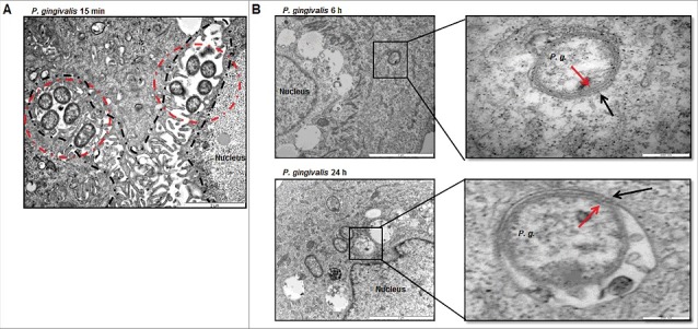
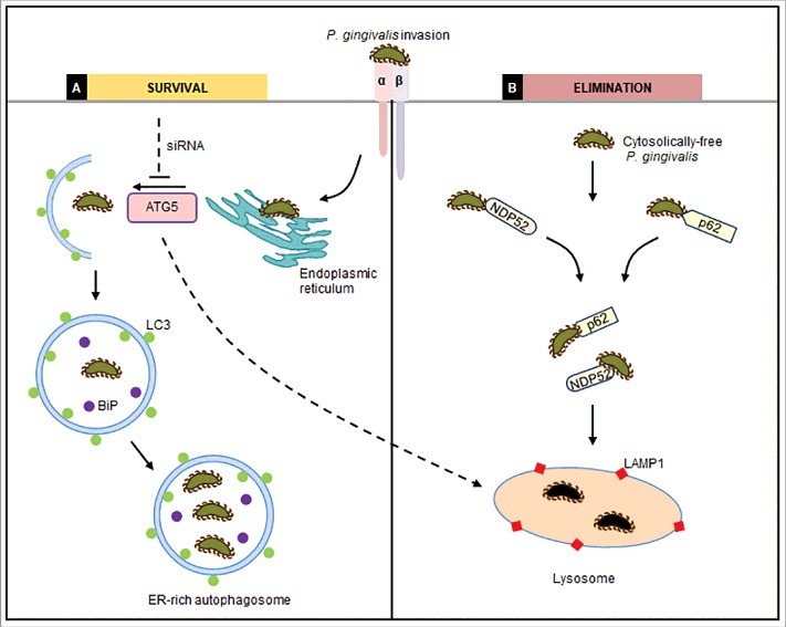
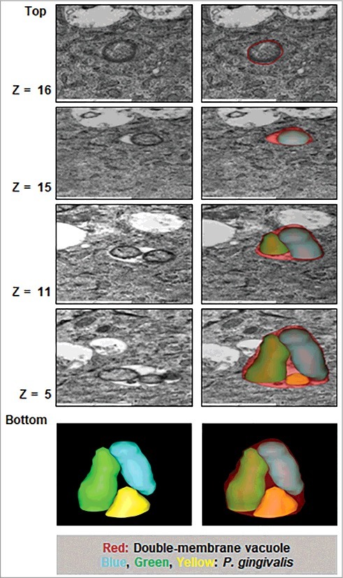
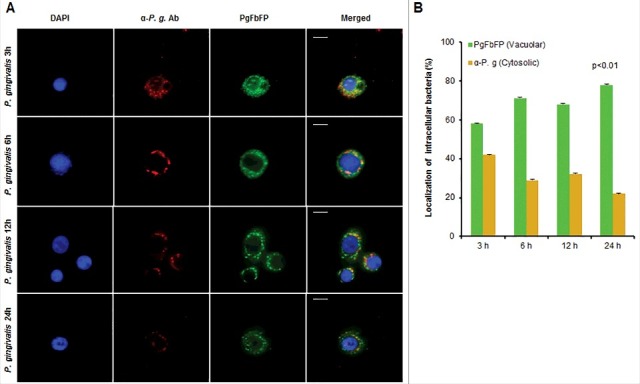
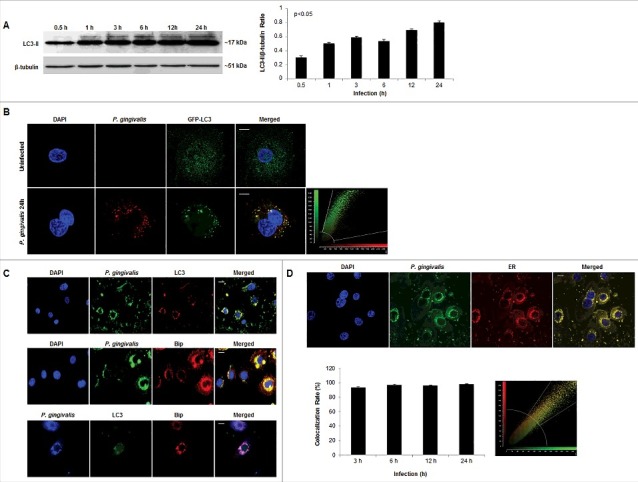
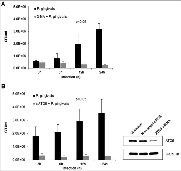
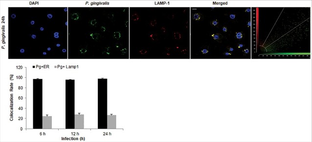
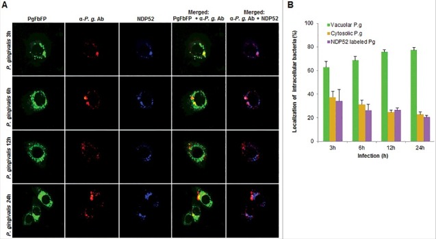
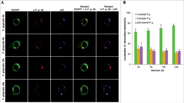
Porphyromonas gingivalis, an opportunistic pathogen usurps gingival epithelial cells (GECs) as primary intracellular niche for its colonization in the oral mucosa. However, the precise characterization of the intracellular trafficking and fate of P. gingivalis in GECs remains incomplete. Therefore, we employed high-resolution three-dimensional-transmission-electron-microscopy to determine the subcellular location of P. gingivalis in human primary GECs upon invasion. Serial sections of infected-GECs and their tomographic reconstruction depicted ER-rich-double-membrane autophagosomal-vacuoles harboring P. gingivalis. Western-blotting and fluorescence confocal microscopy showed that P. gingivalis significantly induces LC3-lipidation in a time-dependent-manner and co-localizes with LC3, ER-lumen-protein Bip, or ER-tracker, which are major components of the phagophore membrane. Furthermore, GECs that were infected with FMN-green-fluorescent transformant-strain (PgFbFP) and selectively permeabilized by digitonin showed rapidly increasing large numbers of double-membrane-vacuolar-P. gingivalis over 24 hours of infection with a low-ratio of cytosolically free-bacteria. Moreover, inhibition of autophagy using 3-methyladenine or ATG5 siRNA significantly reduced the viability of intracellular P. gingivalis in GECs as determined by an antibiotic-protection-assay. Lysosomal marker, LAMP-1, showed a low-degree colocalization with P. gingivalis (∼20%). PgFbFP was used to investigate the fate of vacuolar- versus cytosolic-P. gingivalis by their association with ubiquitin-binding-adaptor-proteins, NDP52 and p62. Only cytosolic-P. gingivalis had a significant association with both markers, which suggests cytosolically-free bacteria are likely destined to the lysosomal-degradation pathway whereas the vacuolar-P. gingivalis survives. Therefore, the results reveal a novel mechanism for P. gingivalis survival in GECs by harnessing host autophagy machinery to establish a successful replicative niche and persistence in the oral mucosa.
牙龈卟啉单胞菌(Porphyromonas gingivalis)是一种机会性病原体,它会侵占牙龈上皮细胞(gingival epithelial cells,GECs)作为其在口腔黏膜定植的主要细胞内小生境。然而,牙龈卟啉单胞菌在 GEC 中的细胞内运输和命运的确切特征尚不完全清楚。因此,我们采用高分辨率三维透射电子显微镜来确定感染后牙龈卟啉单胞菌在人原代 GEC 中的亚细胞位置。感染 GEC 的连续切片及其断层重建图显示富含内质网的双层膜自噬小泡中含有牙龈卟啉单胞菌。Western blot 和荧光共聚焦显微镜显示,牙龈卟啉单胞菌在时间依赖性方式下显著诱导 LC3 脂质化,并与 LC3、内质网腔蛋白 Bip 或内质网追踪剂共定位,这些都是噬泡膜的主要成分。此外,用 FMN 绿色荧光转化株(PgFbFP)感染的 GEC 并用鱼精蛋白选择性通透后,在 24 小时的感染过程中,迅速增加了大量双层膜囊泡中的牙龈卟啉单胞菌,而细胞质中游离细菌的比例较低。此外,用 3-甲基腺嘌呤或 ATG5 siRNA 抑制自噬,通过抗生素保护试验显著降低了 GEC 中细胞内牙龈卟啉单胞菌的活力。溶酶体标记物 LAMP-1 与牙龈卟啉单胞菌的共定位程度较低(约 20%)。用 PgFbFP 研究囊泡内和细胞质内牙龈卟啉单胞菌的命运,通过与泛素结合衔接蛋白 NDP52 和 p62 结合。只有细胞质内的牙龈卟啉单胞菌与这两种标志物有显著的关联,这表明细胞质内的游离细菌可能注定要进入溶酶体降解途径,而囊泡内的牙龈卟啉单胞菌则存活下来。因此,这些结果揭示了牙龈卟啉单胞菌在 GEC 中通过利用宿主自噬机制建立成功的复制小生境和在口腔黏膜中持续存在的新机制。