Zhang Dongzhe, Qu Junwei, Ke Cuncun, Kong Xiumei, Liu Mengyun, Nawaz Khan Iqbal, Huang Shuxin, Tian Haijiao, Xie Tong, Qiu Ke, Li Jing, Wang Mingli, Li Hui, Yuan Fengling, Guo Weikai, Cao Mingya, Zhang Jing, Zhu Keke, Luo Jin, Zhang Fengyan, Cui Xiukun, Mu Hongmei, Hu Yanzhong
Division of Vision Science, Joint National Laboratory for Antibody Drug Engineering, Henan University, Kaifeng, China.
Kaifeng Key Lab for Cataract and Myopia, Institute of Eye Disease, Kaifeng Central Hospital, Kaifeng, China.
Invest Ophthalmol Vis Sci. 2025 Apr 1;66(4):68. doi: 10.1167/iovs.66.4.68.
We studied the regulatory association of Porphyromonas gingivalis (PG) and cataracts.
PCR and FISH assays were used for detecting PG 16s ribosomal RNA genome, Immunofluorescence was for expression of RpgA in anterior capsular epithelium and fibrosis markers in anterior subcapsular cataract (ASC) model. Flow cytometry was for reactive oxygen species and apoptosis. RNA deep sequencing is for differential gene expression analysis.
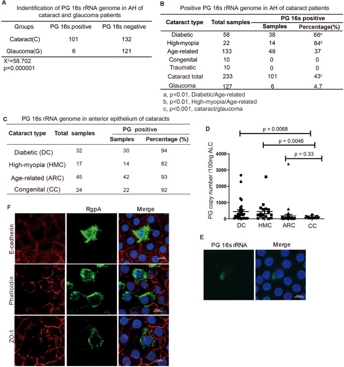
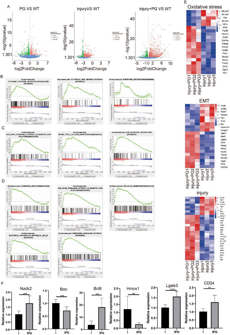
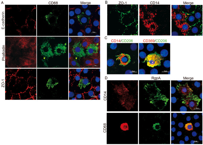
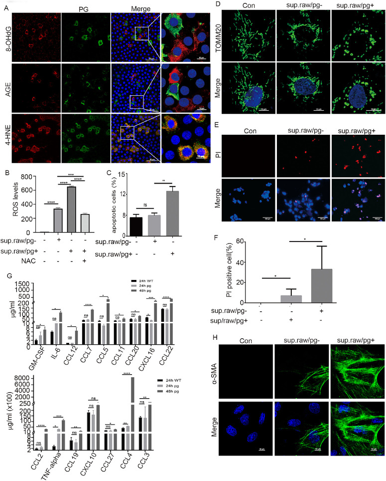
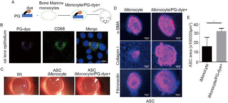
PG's 16s ribosomal RNA gene is positively in 43.3% (101/233 cases) of aqueous humor (AH) samples of patients with cataracts, which differs from 4.7% (6/127) of PG-positive AH in patients with glaucoma. Diabetic and high myopia cataracts increase PG-positive AH compared with age-related cataracts. No PG is observed in AH of congenital cataracts. PG is positive in 82% to 94% of the cataractous anterior capsule tissues from high myopia and age-related, congenital, and diabetic cataracts. The PG-positive cells in the cataractous anterior capsular epithelium are CD68+/CD14+ macrophages, but not anterior epithelial cells. In rat ASC models, PG injected via the tail vein or PG-carried bone marrow monocytes can migrate into the equatorial lens epithelium in form of PG-positive macrophages, which promote ASC progression with upregulation of collagen, fibronectin and α smooth muscle actin (α-SMA) expression, and increase 8-OHdG levels and α-SMA expression in the surrounding lens epithelial cells. Kyoto Encyclopedia of Genes and Genomes and Gene Ontology analysis of the RNA sequencing dataset of ASC tissues shows that signaling pathways related to epithelial-mesenchymal transition, oxidative stress, and cell death are up-regulated in PG + ASC compared with that in ASC alone. Co-culture of supernatants of Raw264.7/PG+ cells with rat primary lens epithelial cells increases the 8-OHdG levels, mitochondrial fission, apoptosis, and expression of α-SMA.
Chronic infection with PG can access the lens epithelium via macrophages during stress conditions, which promotes cataract development by possibly elevating oxidative stress, apoptosis, and epithelial-mesenchymal transition in lens tissues. PG infection is a novel a risk factor for cataract development.
我们研究了牙龈卟啉单胞菌(PG)与白内障之间的调控关联。
采用聚合酶链反应(PCR)和荧光原位杂交(FISH)检测PG 16S核糖体RNA基因组,免疫荧光法检测前囊上皮中RpgA的表达以及前囊下白内障(ASC)模型中的纤维化标志物。流式细胞术检测活性氧和细胞凋亡。RNA深度测序用于差异基因表达分析。
白内障患者房水(AH)样本中43.3%(101/233例)的PG 16S核糖体RNA基因呈阳性,这与青光眼患者中4.7%(6/127)的PG阳性AH不同。与年龄相关性白内障相比,糖尿病性和高度近视性白内障患者的PG阳性AH增加。先天性白内障患者的AH中未观察到PG。高度近视性、年龄相关性、先天性和糖尿病性白内障的白内障前囊组织中82%至94%的PG呈阳性。白内障前囊上皮中的PG阳性细胞是CD68+/CD14+巨噬细胞,而非前上皮细胞。在大鼠ASC模型中,经尾静脉注射的PG或携带PG的骨髓单核细胞可作为PG阳性巨噬细胞迁移至晶状体赤道部上皮,通过上调胶原蛋白、纤连蛋白和α平滑肌肌动蛋白(α-SMA)的表达促进ASC进展,并增加周围晶状体上皮细胞中的8-羟基脱氧鸟苷(8-OHdG)水平和α-SMA表达。对ASC组织的RNA测序数据集进行京都基因与基因组百科全书(KEGG)和基因本体论(GO)分析表明,与单独的ASC相比,PG + ASC中与上皮-间质转化、氧化应激和细胞死亡相关的信号通路上调。将Raw264.7/PG+细胞的上清液与大鼠原代晶状体上皮细胞共培养可增加8-OHdG水平、线粒体裂变、细胞凋亡以及α-SMA的表达。
PG的慢性感染在应激条件下可通过巨噬细胞进入晶状体上皮,可能通过提高晶状体组织中的氧化应激、细胞凋亡和上皮-间质转化促进白内障发展。PG感染是白内障发生发展的一个新的危险因素。