Orthopaedic Department, The First Affiliated Hospital of Nanjing Medical University, Nanjing, Jiangsu 210029, P.R. China.
Orthopaedic Department, The First Affiliated Hospital of Nanjing Medical University, Nanjing, Jiangsu 210029, P.R. China.
Int J Mol Med. 2018 Jul;42(1):171-181. doi: 10.3892/ijmm.2018.3614. Epub 2018 Apr 3.
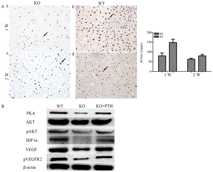
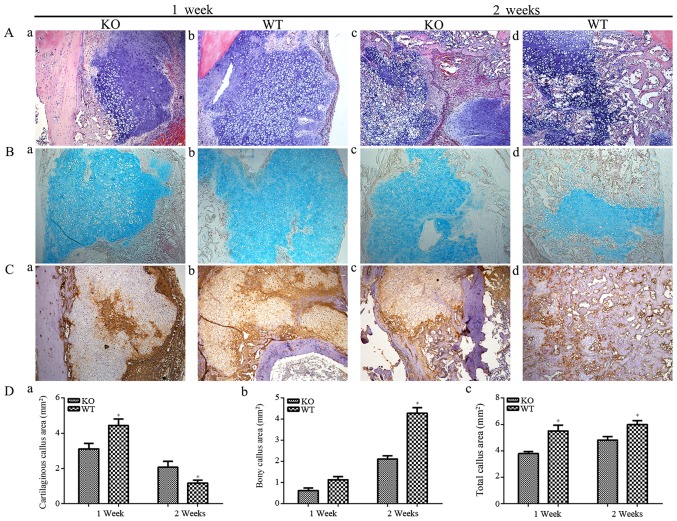
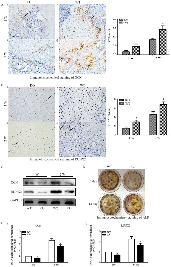
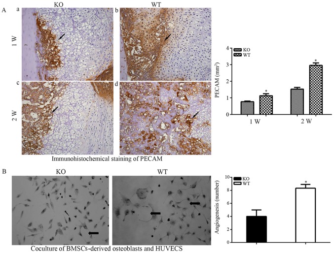
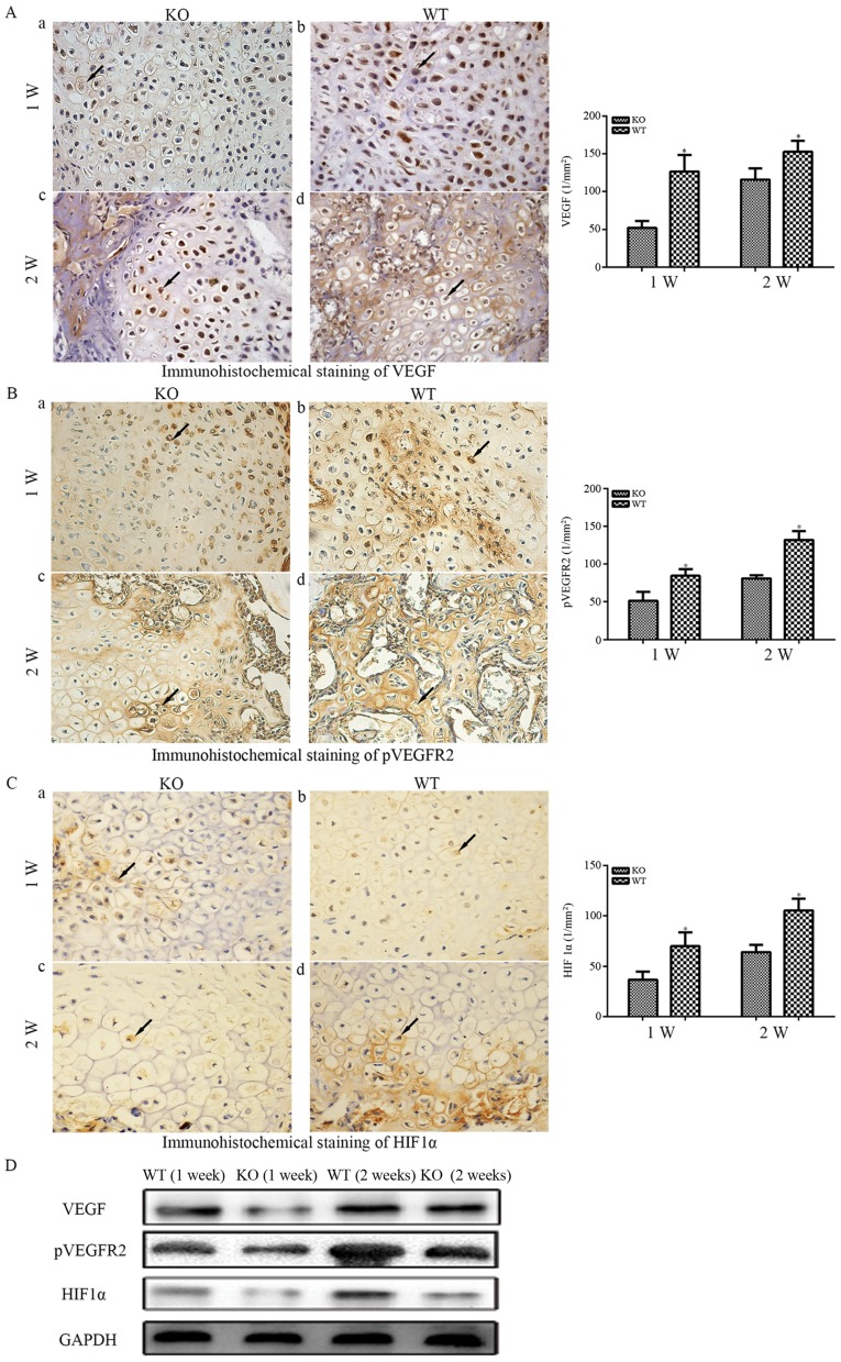
Intermittent low‑dose injections of parathyroid hormone (PTH) have been reported to exert bone anabolic effects and to promote fracture healing. As an important proangiogenic cytokine, vascular endothelial growth factor (VEGF) is secreted by bone marrow mesenchymal stem cells (BMSCs) and osteoblasts, and serves a crucial regulatory role in the process of vascular development and regeneration. To investigate whether lack of endogenous PTH causes reduced angiogenic capacity and thereby delays the process of fracture healing by downregulating the VEGF signaling pathway, a PTH knockout (PTHKO) mouse fracture model was generated. Fracture healing was observed using X‑ray and micro‑computerized tomography. Bone anabolic and angiogenic markers were analyzed by immunohistochemistry and western blot analysis. The expression levels of VEGF and associated signaling pathways in murine BMSC‑derived osteoblasts were measured by quantitative polymerase chain reaction and western blot analysis. The expression levels of protein kinase A (PKA), phosphorylated‑serine/threonine protein kinase (pAKT), hypoxia‑inducible factor‑1α (HIF1α) and VEGF were significantly decreased in BMSC‑derived osteoblasts from PTHKO mice. In addition, positive platelet endothelial cell adhesion molecule staining was reduced in PTHKO mice, as determined by immunohistochemistry. The expression levels of HIF1α, VEGF, runt‑related transcription factor 2, osteocalcin and alkaline phosphatase were also decreased in PTHKO mice, and fracture healing was delayed. In conclusion, lack of endogenous PTH may reduce VEGF expression in BMSC‑derived osteoblasts by downregulating the activity of the PKA/pAKT/HIF1α/VEGF pathway, thus affecting endochondral bone formation by causing a reduction in angiogenesis and osteogenesis, ultimately leading to delayed fracture healing.
甲状旁腺激素(PTH)间歇性低剂量注射已被报道具有促进骨合成代谢和促进骨折愈合的作用。血管内皮生长因子(VEGF)作为一种重要的促血管生成细胞因子,由骨髓间充质干细胞(BMSCs)和成骨细胞分泌,在血管发育和再生过程中发挥着关键的调节作用。为了研究内源性 PTH 的缺乏是否通过下调 VEGF 信号通路导致血管生成能力降低,从而延迟骨折愈合过程,我们构建了 PTH 敲除(PTHKO)小鼠骨折模型。通过 X 射线和微计算机断层扫描观察骨折愈合情况。通过免疫组织化学和 Western blot 分析检测骨合成和血管生成标志物。通过定量聚合酶链反应和 Western blot 分析测量小鼠 BMSC 源性成骨细胞中 VEGF 及其相关信号通路的表达水平。PKA、磷酸化丝氨酸/苏氨酸蛋白激酶(pAKT)、缺氧诱导因子-1α(HIF1α)和 VEGF 的蛋白表达水平在 PTHKO 小鼠的 BMSC 源性成骨细胞中显著降低。此外,通过免疫组织化学分析,PTHKO 小鼠的血小板内皮细胞黏附分子阳性染色减少。在 PTHKO 小鼠中,HIF1α、VEGF、Runt 相关转录因子 2、骨钙素和碱性磷酸酶的表达水平也降低,骨折愈合延迟。总之,内源性 PTH 的缺乏可能通过下调 PKA/pAKT/HIF1α/VEGF 通路的活性降低 BMSC 源性成骨细胞中 VEGF 的表达,从而通过减少血管生成和成骨作用影响软骨内骨形成,最终导致骨折愈合延迟。