Witt Benjamin L, Factor Rachel E, Chadwick Barbara E, Caron Justin, Siddiqui Ali A, Adler Douglas G
Department of Pathology, School of Medicine, ARUP Laboratories, University of Utah, Salt Lake City, UT, USA.
Division of Gastroenterology, School of Medicine, Thomas Jefferson University, Philadelphia, PA, USA.
Endosc Ultrasound. 2018 Sep-Oct;7(5):323-328. doi: 10.4103/eus.eus_51_17.
EUS guided core biopsy was once rarely performed but is now entering mainstream practice. Neuroendocrine tumors often warrant core biopsy as sufficient tissue must be obtained to allow for special staining to ensure a correct diagnosis. Traditionally these lesions were sampled with FNA needles. We performed a retrospective pilot study to evaluate the clinical value and efficacy of the a new EUS core needle biopsy needle as compared to a standard EUS FNA needle in the evaluation of patients with known or suspected neuroendocrine tumors.
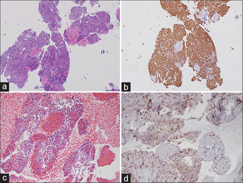
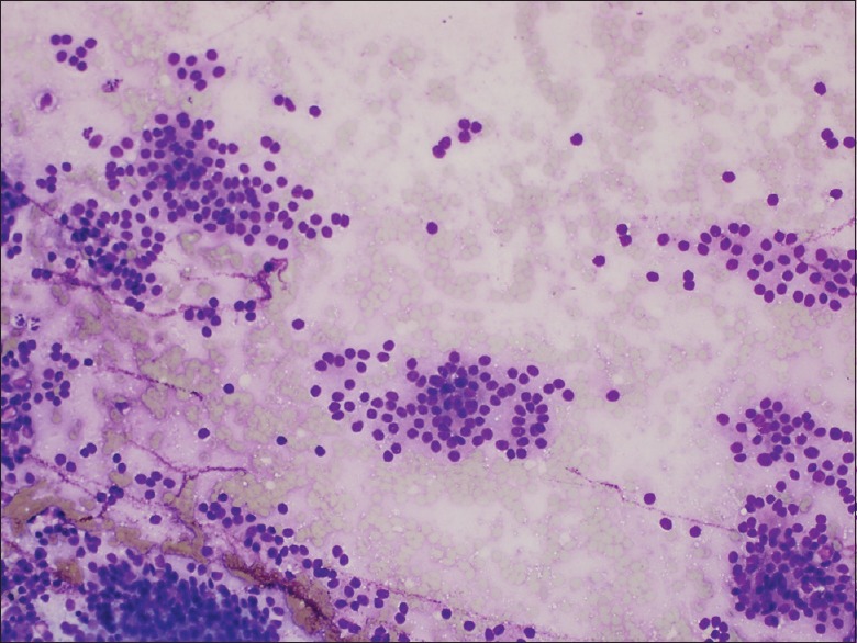
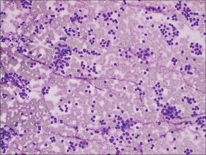
A retrospective analysis of the first 10 patients (between January 2015 and April 2016) to undergo EUS-FNA with the SharkCore needle at the University of Utah School of Medicine/Huntsman Cancer Center with neuroendocrine tumors. Each case was retrospectively reviewed by a board certified cytopathologist (BLW) for the following cytologic parameters on the aspirate smears or touch/squash preparations: overall cellularity [1 (low) to 3 (high)], percentage of obtained cells that were lesional/representative (<25%, 26%-50%, and >50%), relative ease of interpretation [1 (difficult) to 3 (easy)]. Pathologic material and reporting records were also reviewed for each case to confirm the number of needle passes to achieve diagnostic adequacy, the presence or absence diagnostic material on H&E slide (from cell block, if prepared), whether a definitive diagnosis was able to be rendered, and the presence or absence of a true core/core fragments (within the cell block, if prepared).
A total of 20 patients underwent EUS-FNA for suspected neuroendocrine lesions. Ten patients underwent either transgastric or transduodenal EUS-FNA with the 22 gauge SharkCore needle. The comparison cohort of 10 patients underwent either transgastric or transduodenal EUS-FNA with the standard 22 gauge Echotip needle. The SharkCore needle required a fewer mean number of needle passes to obtain diagnostic adequacy than the Echotip (P=0.0074). For cases with cell blocks, the SharkCore needle produced diagnostic material in 100% of cases, whereas Echotip produced diagnostic material in 60% of cases. There was no significant difference between specimen cellularity, percentage of lesional material, or ease of interpretation between the two needle types.
Our pilot investigation targeting patients with known or suspected pancreatic NETs indicates that the SharkCore needle shows promise in obtaining suitable tissue for ancillary testing that can allow for more definitive pathologic interpretations on EUS FNA specimens. Fewer passes were needed with the core needle when compared to a standard needle.
超声内镜引导下的核心活检术曾很少实施,但如今正逐渐成为主流操作。神经内分泌肿瘤常常需要进行核心活检,因为必须获取足够的组织以进行特殊染色,从而确保做出正确诊断。传统上,这些病变是用细针穿刺抽吸(FNA)针进行采样的。我们进行了一项回顾性试点研究,以评估一种新型超声内镜核心活检针与标准超声内镜FNA针相比,在评估已知或疑似神经内分泌肿瘤患者时的临床价值和有效性。
对犹他大学医学院/亨茨曼癌症中心首批10例(2015年1月至2016年4月)接受使用SharkCore针进行超声内镜FNA检查的神经内分泌肿瘤患者进行回顾性分析。每例病例均由一位经过委员会认证的细胞病理学家(BLW)对吸取涂片或触摸/压片标本进行回顾性检查,以获取以下细胞学参数:总体细胞含量[1(低)至3(高)]、获取的病变/代表性细胞的百分比(<25%、26%-50%和>50%)、相对易解释性[1(困难)至3(容易)]。还对每个病例的病理材料和报告记录进行了审查,以确认达到诊断充分性所需的穿刺次数、苏木精-伊红(H&E)玻片(如制备了细胞块)上是否存在诊断材料、是否能够做出明确诊断以及是否存在真正的核心/核心碎片(如制备了细胞块)。
共有20例患者因疑似神经内分泌病变接受了超声内镜FNA检查。10例患者使用22号SharkCore针进行了经胃或经十二指肠超声内镜FNA检查。10例患者的对照队列使用标准22号Echotip针进行了经胃或经十二指肠超声内镜FNA检查。与Echotip针相比,SharkCore针获得诊断充分性所需的平均穿刺次数更少(P=0.0074)。对于有细胞块的病例,SharkCore针在100%的病例中产生了诊断材料,而Echotip针在60%的病例中产生了诊断材料。两种针型在标本细胞含量、病变材料百分比或易解释性方面没有显著差异。
我们针对已知或疑似胰腺神经内分泌肿瘤患者的试点研究表明,SharkCore针在获取适合辅助检测的组织方面显示出前景,这可以使超声内镜FNA标本的病理解释更加明确。与标准针相比,核心针所需的穿刺次数更少。