Omami Galal
American Board of Oral and Maxillofacial Radiology, United States.
University of Kentucky College of Dentistry, Department of Oral Health Practice, Division of Oral Medicine, Diagnosis and Radiology, 800 Rose Street, Room MN-320, Lexington, KY 40536-0297, United States.
Saudi Dent J. 2018 Apr;30(2):151-154. doi: 10.1016/j.sdentj.2017.12.003. Epub 2017 Dec 24.
To investigate the prevalence and pattern of calcification of the stylohyoid complex in Libyan population.
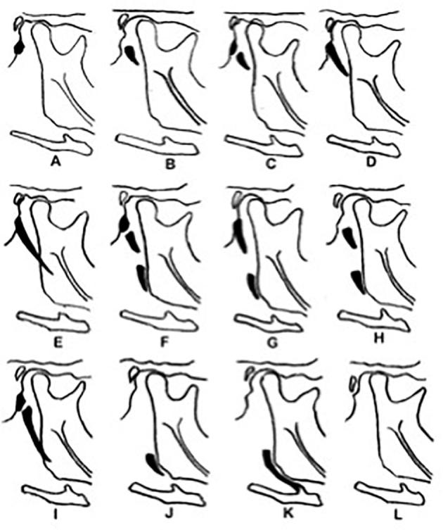
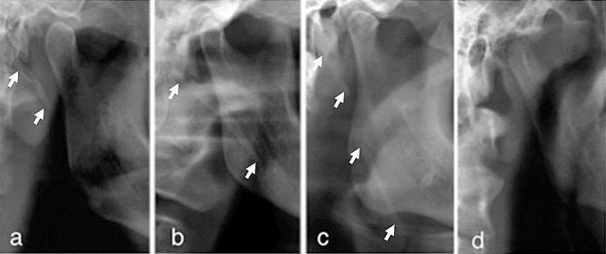
Archived digital panoramic radiographs of 3343 patients were collected; 181 images were excluded for underage or poor image quality. Thus, the images of 3162 patients (1081 men, 2081 women; women-to-men ratio, 2:1; age range, 16-68 years; mean age, 36.7 years) retrieved and assigned to one of four morphological patterns of the stylohyoid complex: regular, elongated, calcified, and undetected. Data were analyzed with the Χ test using SPSS (Chicago, IL, USA); P values lower than 0.05 were considered statistically significant.
Out of 3162 images studied, the styloid process was demonstrated to be regular in 1935 (61.2%), elongated in 541 (17.2%), calcified in 565 (17.8%), and undetected in 121 (3.8%). Symmetric patterns were demonstrated on 2580 (81.6%) images. An elongated stylohyoid complex was significantly more common in women than in men (P = .0404).
The anatomical patterns of the stylohyoid complex in Libyans were highly variable. Dental clinicians should recognize the various morphological patterns of the stylohyoid complex on panoramic radiographs. Computed tomography studies are recommended for further morphometric analysis of the stylohyoid complex.
调查利比亚人群茎突舌骨复合体钙化的发生率及模式。
收集3343例患者的数字化全景X线片存档资料;181张图像因患者未成年或图像质量差被排除。因此,获取了3162例患者(1081名男性,2081名女性;女性与男性比例为2:1;年龄范围16 - 68岁;平均年龄36.7岁)的图像,并将其分为茎突舌骨复合体的四种形态模式之一:规则型、细长型、钙化型和未检出型。使用SPSS(美国伊利诺伊州芝加哥)进行χ检验分析数据;P值低于0.05被认为具有统计学意义。
在研究的3162张图像中,茎突显示为规则型的有1935张(61.2%),细长型的有541张(17.2%),钙化型的有565张(17.8%),未检出的有121张(3.8%)。2580张(81.6%)图像显示为对称模式。茎突舌骨复合体细长型在女性中比男性更常见(P = 0.0404)。
利比亚人茎突舌骨复合体的解剖模式差异很大。牙科临床医生应在全景X线片上识别茎突舌骨复合体的各种形态模式。建议进行计算机断层扫描研究以进一步对茎突舌骨复合体进行形态测量分析。