• 文献检索
  • 文档翻译
  • 深度研究
  • 学术资讯
  • Suppr Zotero 插件Zotero 插件
  • 邀请有礼
  • 套餐&价格
  • 历史记录
应用&插件
Suppr Zotero 插件Zotero 插件浏览器插件Mac 客户端Windows 客户端微信小程序
定价
高级版会员购买积分包购买API积分包
服务
文献检索文档翻译深度研究API 文档MCP 服务
关于我们
关于 Suppr公司介绍联系我们用户协议隐私条款
关注我们

Suppr 超能文献

核心技术专利:CN118964589B侵权必究
粤ICP备2023148730 号-1Suppr @ 2026

文献检索

告别复杂PubMed语法,用中文像聊天一样搜索,搜遍4000万医学文献。AI智能推荐,让科研检索更轻松。

立即免费搜索

文件翻译

保留排版,准确专业,支持PDF/Word/PPT等文件格式,支持 12+语言互译。

免费翻译文档

深度研究

AI帮你快速写综述,25分钟生成高质量综述,智能提取关键信息,辅助科研写作。

立即免费体验

用于早期青光眼诊断的北卡罗来纳大学光学相干断层扫描指数的验证

Validation of the UNC OCT Index for the Diagnosis of Early Glaucoma.

作者信息

Mwanza Jean-Claude, Lee Gary, Budenz Donald L, Warren Joshua L, Wall Michael, Artes Paul H, Callan Thomas M, Flanagan John G

机构信息

Department of Ophthalmology, University of North Carolina at Chapel Hill, Chapel Hill, NC, USA.

Research and Development, Carl Zeiss Meditec, Inc., Dublin, CA, USA.

出版信息

Transl Vis Sci Technol. 2018 Apr 3;7(2):16. doi: 10.1167/tvst.7.2.16. eCollection 2018 Apr.

DOI:10.1167/tvst.7.2.16
PMID:29629238
原文链接:https://pmc.ncbi.nlm.nih.gov/articles/PMC5886105/
Abstract

PURPOSE

To independently validate the performance of the University of North Carolina Optical Coherence Tomography (UNC OCT) Index in diagnosing and predicting early glaucoma.

METHODS

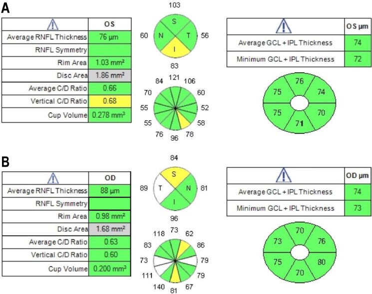
Data of 118 normal subjects (118 eyes) and 96 subjects (96 eyes) with early glaucoma defined as visual field mean deviation (MD) greater than -4 decibels (dB), aged 40 to 80 years, and who were enrolled in the Full-Threshold Testing Size III, V, VI comparison study were used in this study. CIRRUS OCT average and quadrants' retinal nerve fiber layer (RNFL); optic disc vertical cup-to-disc ratio (VCDR), cup-to-disc area ratio, and rim area; and average, minimum, and six sectoral ganglion cell-inner plexiform layer (GCIPL) measurements were run through the UNC OCT Index algorithm. Area under the receiver operating characteristic curve (AUC) and sensitivities at 95% and 99% specificity were calculated and compared between single parameters and the UNC OCT Index.

RESULTS

Mean age was 60.1 ± 11.0 years for normal subjects and 66.5 ± 8.1 years for glaucoma patients ( < 0.001). MD was 0.29 ± 1.04 dB and -1.30 ± 1.35 dB in normal and glaucomatous eyes ( < 0.001), respectively. The AUC of the UNC OCT Index was 0.96. The best single metrics when compared to the UNC OCT Index were VCDR (0.93, = 0.054), average RNFL (0.92, = 0.014), and minimum GCIPL (0.91, = 0.009). The sensitivities at 95% and 99% specificity were 85.4% and 76.0% (UNC OCT Index), 71.9% and 62.5% (VCDR, all < 0.001), 64.6% and 53.1% (average RNFL, all < 0.001), and 66.7% and 58.3% (minimum GCIPL, all < 0.001), respectively.

CONCLUSIONS

The findings confirm that the UNC OCT Index may provide improved diagnostic perforce over that of single OCT parameters and may be a good tool for detection of early glaucoma.

TRANSLATIONAL RELEVANCE

The UNC OCT Index algorithm may be incorporated easily into routine clinical practice and be useful for detecting early glaucoma.

摘要

目的

独立验证北卡罗来纳大学光学相干断层扫描(UNC OCT)指数在诊断和预测早期青光眼方面的性能。

方法

本研究使用了118名正常受试者(118只眼)和96名早期青光眼受试者(96只眼)的数据,这些受试者年龄在40至80岁之间,早期青光眼定义为视野平均偏差(MD)大于-4分贝(dB),并参与了全阈值测试大小III、V、VI比较研究。对CIRRUS OCT平均和象限视网膜神经纤维层(RNFL);视盘垂直杯盘比(VCDR)、杯盘面积比和边缘面积;以及平均、最小和六个扇形神经节细胞-内丛状层(GCIPL)测量值进行UNC OCT指数算法运算。计算并比较单参数与UNC OCT指数之间的受试者操作特征曲线下面积(AUC)以及95%和99%特异性时的敏感性。

结果

正常受试者的平均年龄为60.1±11.0岁,青光眼患者为66.5±8.1岁(P<0.001)。正常眼和青光眼眼中的MD分别为0.29±1.04 dB和-1.30±1.35 dB(P<0.001)。UNC OCT指数的AUC为0.96。与UNC OCT指数相比,最佳的单一指标是VCDR(0.93,P = 0.054)、平均RNFL(0.92,P = 0.014)和最小GCIPL(0.91,P = 0.009)。95%和99%特异性时的敏感性分别为85.4%和76.0%(UNC OCT指数)、71.9%和62.5%(VCDR,均P<0.001)、64.6%和53.1%(平均RNFL,均P<0.001)以及66.7%和58.3%(最小GCIPL,均P<0.001)。

结论

研究结果证实,UNC OCT指数在诊断效能上可能优于单一OCT参数,可能是检测早期青光眼的良好工具。

转化相关性

UNC OCT指数算法可轻松纳入常规临床实践,有助于检测早期青光眼。

https://cdn.ncbi.nlm.nih.gov/pmc/blobs/dafe/5886105/54a489d97683/i2164-2591-7-2-16-f03.jpg
https://cdn.ncbi.nlm.nih.gov/pmc/blobs/dafe/5886105/f44c274c96b5/i2164-2591-7-2-16-f01.jpg
https://cdn.ncbi.nlm.nih.gov/pmc/blobs/dafe/5886105/77e75726d9ca/i2164-2591-7-2-16-f02.jpg
https://cdn.ncbi.nlm.nih.gov/pmc/blobs/dafe/5886105/54a489d97683/i2164-2591-7-2-16-f03.jpg
https://cdn.ncbi.nlm.nih.gov/pmc/blobs/dafe/5886105/f44c274c96b5/i2164-2591-7-2-16-f01.jpg
https://cdn.ncbi.nlm.nih.gov/pmc/blobs/dafe/5886105/77e75726d9ca/i2164-2591-7-2-16-f02.jpg
https://cdn.ncbi.nlm.nih.gov/pmc/blobs/dafe/5886105/54a489d97683/i2164-2591-7-2-16-f03.jpg

相似文献

1
Validation of the UNC OCT Index for the Diagnosis of Early Glaucoma.用于早期青光眼诊断的北卡罗来纳大学光学相干断层扫描指数的验证
Transl Vis Sci Technol. 2018 Apr 3;7(2):16. doi: 10.1167/tvst.7.2.16. eCollection 2018 Apr.
2
Evaluation of University of North Carolina OCT Index for Diagnosis of Early Glaucoma.评价北卡罗来纳大学 OCT 指数对早期青光眼的诊断价值。
Ophthalmol Glaucoma. 2022 Sep-Oct;5(5):490-497. doi: 10.1016/j.ogla.2022.03.001. Epub 2022 Mar 8.
3
Diagnostic accuracy of macular ganglion cell-inner plexiform layer thickness for glaucoma detection in a population-based study: Comparison with optic nerve head imaging parameters.基于人群的研究中,黄斑神经节细胞-内丛状层厚度对青光眼检测的诊断准确性:与视神经头成像参数的比较。
PLoS One. 2018 Jun 26;13(6):e0199134. doi: 10.1371/journal.pone.0199134. eCollection 2018.
4
Ability of cirrus HD-OCT optic nerve head parameters to discriminate normal from glaucomatous eyes.Cirrus HD-OCT视神经乳头参数区分正常眼与青光眼性眼的能力。
Ophthalmology. 2011 Feb;118(2):241-8.e1. doi: 10.1016/j.ophtha.2010.06.036. Epub 2010 Oct 28.
5
Macular ganglion cell/inner plexiform layer measurements by spectral domain optical coherence tomography for detection of early glaucoma and comparison to retinal nerve fiber layer measurements.利用谱域光学相干断层扫描测量黄斑神经节细胞/内丛状层,以早期检测青光眼,并与视网膜神经纤维层测量值进行比较。
Am J Ophthalmol. 2013 Dec;156(6):1297-1307.e2. doi: 10.1016/j.ajo.2013.08.001. Epub 2013 Sep 25.
6
A Diagnostic Calculator for Detecting Glaucoma on the Basis of Retinal Nerve Fiber Layer, Optic Disc, and Retinal Ganglion Cell Analysis by Optical Coherence Tomography.一种基于光学相干断层扫描技术对视网膜神经纤维层、视盘和视网膜神经节细胞进行分析来检测青光眼的诊断计算器。
Invest Ophthalmol Vis Sci. 2015 Oct;56(11):6788-95. doi: 10.1167/iovs.15-17176.
7
[Significance of optic disc topography and retinal nerve fiber layer thickness measurement by spectral-domain OCT in diagnosis of glaucoma].[频域光学相干断层扫描测量视盘地形图及视网膜神经纤维层厚度在青光眼诊断中的意义]
Zhonghua Yan Ke Za Zhi. 2010 Aug;46(8):702-8.
8
Glaucoma diagnostic accuracy of optical coherence tomography parameters in early glaucoma with different types of optic disc damage.光学相干断层扫描参数对不同类型视盘损伤早期青光眼的诊断准确性。
Ophthalmology. 2014 Oct;121(10):1990-7. doi: 10.1016/j.ophtha.2014.04.030. Epub 2014 Jun 14.
9
Glaucoma diagnostic accuracy of ganglion cell-inner plexiform layer thickness: comparison with nerve fiber layer and optic nerve head.节细胞-内丛状层厚度对青光眼的诊断准确性:与神经纤维层和视神经盘的比较。
Ophthalmology. 2012 Jun;119(6):1151-8. doi: 10.1016/j.ophtha.2011.12.014. Epub 2012 Feb 23.
10
Ganglion cell-inner plexiform layer thickness of high definition optical coherence tomography in perimetric and preperimetric glaucoma.高分辨率光学相干断层扫描在视野缺损性青光眼和早期青光眼患者中的神经节细胞-内丛状层厚度研究
Invest Ophthalmol Vis Sci. 2014 Jul 11;55(8):4768-75. doi: 10.1167/iovs.14-14598.

引用本文的文献

1
A Pattern-Based OCT Metric for Glaucoma Detection.一种基于模式的用于青光眼检测的光学相干断层扫描指标。
Transl Vis Sci Technol. 2024 Dec 2;13(12):21. doi: 10.1167/tvst.13.12.21.
2
Independent Effects of Blood Pressure on Intraocular Pressure and Retinal Ganglion Cell Degeneration: A Mendelian Randomization Study.血压对眼压和视网膜神经节细胞变性影响的独立作用:一项孟德尔随机研究。
Invest Ophthalmol Vis Sci. 2024 Jul 1;65(8):35. doi: 10.1167/iovs.65.8.35.
3
Unveiling Novel Structural Biomarkers for the Diagnosis of Glaucoma.揭示用于青光眼诊断的新型结构生物标志物。

本文引用的文献

1
Applicability of automatic spectral domain optical coherence tomography for glaucoma mass screening.自动谱域光学相干断层扫描在青光眼群体筛查中的适用性。
Clin Ophthalmol. 2016 Dec 29;11:97-103. doi: 10.2147/OPTH.S115547. eCollection 2017.
2
Green disease in optical coherence tomography diagnosis of glaucoma.光学相干断层扫描在青光眼诊断中的绿色疾病
Curr Opin Ophthalmol. 2017 Mar;28(2):139-153. doi: 10.1097/ICU.0000000000000353.
3
Risk of Visual Field Progression in Glaucoma Patients with Progressive Retinal Nerve Fiber Layer Thinning: A 5-Year Prospective Study.
Biomedicines. 2024 May 29;12(6):1211. doi: 10.3390/biomedicines12061211.
4
Performance evaluation of various deep learning based models for effective glaucoma evaluation using optical coherence tomography images.使用光学相干断层扫描图像对各种基于深度学习的模型进行有效青光眼评估的性能评估。
Multimed Tools Appl. 2022;81(19):27737-27781. doi: 10.1007/s11042-022-12826-y. Epub 2022 Mar 28.
5
A Case for the Use of Artificial Intelligence in Glaucoma Assessment.人工智能在青光眼评估中的应用案例。
Ophthalmol Glaucoma. 2022 May-Jun;5(3):e3-e13. doi: 10.1016/j.ogla.2021.12.003. Epub 2021 Dec 22.
6
A Simple Subjective Evaluation of Enface OCT Reflectance Images Distinguishes Glaucoma From Healthy Eyes.正视 OCT 反射图像的简单主观评估可区分青光眼与健康眼。
Transl Vis Sci Technol. 2021 May 3;10(6):31. doi: 10.1167/tvst.10.6.31.
7
A longitudinal comparison in cynomolgus macaques of the effect of brimonidine on optic nerve neuropathy using diffusion tensor imaging magnetic resonance imaging and spectral domain optical coherence tomography.使用扩散张量成像磁共振成像和光谱域光学相干断层扫描对食蟹猴进行溴莫尼定对视神经病变影响的纵向比较。
Heliyon. 2021 Apr 6;7(4):e06701. doi: 10.1016/j.heliyon.2021.e06701. eCollection 2021 Apr.
8
High interpretable machine learning classifier for early glaucoma diagnosis.用于早期青光眼诊断的高可解释性机器学习分类器。
Int J Ophthalmol. 2021 Mar 18;14(3):393-398. doi: 10.18240/ijo.2021.03.10. eCollection 2021.
9
Evaluation of the external validity of a joint structure-function model for monitoring glaucoma progression.评估用于监测青光眼进展的联合结构-功能模型的外部有效性。
Sci Rep. 2020 Nov 12;10(1):19701. doi: 10.1038/s41598-020-76834-4.
10
Macular imaging with optical coherence tomography in glaucoma.光学相干断层扫描在青光眼中的黄斑成像。
Surv Ophthalmol. 2020 Nov-Dec;65(6):597-638. doi: 10.1016/j.survophthal.2020.03.002. Epub 2020 Mar 19.
青光眼患者视网膜神经纤维层进行性变薄的视野进展风险:一项 5 年前瞻性研究。
Ophthalmology. 2016 Jun;123(6):1201-10. doi: 10.1016/j.ophtha.2016.02.017. Epub 2016 Mar 19.
4
Predicting Development of Glaucomatous Visual Field Conversion Using Baseline Fourier-Domain Optical Coherence Tomography.利用基线傅里叶域光学相干断层扫描预测青光眼视野转换的发展
Am J Ophthalmol. 2016 Mar;163:29-37. doi: 10.1016/j.ajo.2015.11.029. Epub 2015 Nov 26.
5
A Diagnostic Calculator for Detecting Glaucoma on the Basis of Retinal Nerve Fiber Layer, Optic Disc, and Retinal Ganglion Cell Analysis by Optical Coherence Tomography.一种基于光学相干断层扫描技术对视网膜神经纤维层、视盘和视网膜神经节细胞进行分析来检测青光眼的诊断计算器。
Invest Ophthalmol Vis Sci. 2015 Oct;56(11):6788-95. doi: 10.1167/iovs.15-17176.
6
Combining measurements from three anatomical areas for glaucoma diagnosis using Fourier-domain optical coherence tomography.使用傅里叶域光学相干断层扫描技术,结合三个解剖区域的测量值进行青光眼诊断。
Br J Ophthalmol. 2015 Sep;99(9):1224-9. doi: 10.1136/bjophthalmol-2014-305907. Epub 2015 Mar 20.
7
Diagnostic classification of macular ganglion cell and retinal nerve fiber layer analysis: differentiation of false-positives from glaucoma.黄斑神经节细胞和视网膜神经纤维层分析的诊断分类:从青光眼区分假阳性。
Ophthalmology. 2015 Mar;122(3):502-10. doi: 10.1016/j.ophtha.2014.09.031. Epub 2014 Nov 14.
8
Discriminating between glaucoma and normal eyes using optical coherence tomography and the 'Random Forests' classifier.使用光学相干断层扫描和“随机森林”分类器区分青光眼和正常眼睛。
PLoS One. 2014 Aug 28;9(8):e106117. doi: 10.1371/journal.pone.0106117. eCollection 2014.
9
Macular ganglion cell analysis for early detection of glaucoma.黄斑神经节细胞分析用于青光眼的早期检测。
Ophthalmology. 2014 Aug;121(8):1508-15. doi: 10.1016/j.ophtha.2014.02.019. Epub 2014 Apr 3.
10
Diagnostic performance of optical coherence tomography ganglion cell--inner plexiform layer thickness measurements in early glaucoma.光学相干断层扫描神经节细胞-内丛状层厚度测量在早期青光眼诊断中的性能。
Ophthalmology. 2014 Apr;121(4):849-54. doi: 10.1016/j.ophtha.2013.10.044. Epub 2014 Jan 3.