• 文献检索
  • 文档翻译
  • 深度研究
  • 学术资讯
  • Suppr Zotero 插件Zotero 插件
  • 邀请有礼
  • 套餐&价格
  • 历史记录
应用&插件
Suppr Zotero 插件Zotero 插件浏览器插件Mac 客户端Windows 客户端微信小程序
定价
高级版会员购买积分包购买API积分包
服务
文献检索文档翻译深度研究API 文档MCP 服务
关于我们
关于 Suppr公司介绍联系我们用户协议隐私条款
关注我们

Suppr 超能文献

核心技术专利:CN118964589B侵权必究
粤ICP备2023148730 号-1Suppr @ 2026

文献检索

告别复杂PubMed语法,用中文像聊天一样搜索,搜遍4000万医学文献。AI智能推荐,让科研检索更轻松。

立即免费搜索

文件翻译

保留排版,准确专业,支持PDF/Word/PPT等文件格式,支持 12+语言互译。

免费翻译文档

深度研究

AI帮你快速写综述,25分钟生成高质量综述,智能提取关键信息,辅助科研写作。

立即免费体验

正视 OCT 反射图像的简单主观评估可区分青光眼与健康眼。

A Simple Subjective Evaluation of Enface OCT Reflectance Images Distinguishes Glaucoma From Healthy Eyes.

机构信息

School of Optometry and Vision Science, University of Bradford, UK.

Ophthalmology Department, Leeds Teaching Hospitals NHS Trust, Leeds, UK.

出版信息

Transl Vis Sci Technol. 2021 May 3;10(6):31. doi: 10.1167/tvst.10.6.31.

DOI:10.1167/tvst.10.6.31
PMID:34036303
原文链接:https://pmc.ncbi.nlm.nih.gov/articles/PMC8161697/
Abstract

PURPOSE

We present a subjective approach to detecting glaucomatous defects in enface images and assess its diagnostic performance. We also test the hypothesis that if reflectivity changes precede thickness changes in glaucoma there should be reduced correlation between the modalities in glaucoma compared to controls.

METHODS

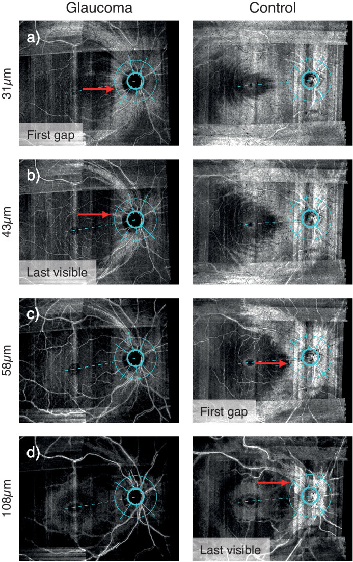
Twenty glaucoma participants and 20 age-matched controls underwent high-resolution OCT scans of one eye. 4 µm-thick enface slabs were constructed through the retina. Enface indices were depths of first gap in visible retinal nerve fiber bundles (RNFBs) and last visible bundle, subjectively evaluated in six sectors of a 3.5 mm circle around the optic disc. Retinal nerve fiber layer thickness (RNFLT) along the same circle was extracted at angles corresponding to enface indices. Between-group differences were tested by linear mixed models. Diagnostic performance was measured by partial receiver operating characteristic area (pAUC).

RESULTS

First gap and last visible bundle were closer to the inner limiting membrane in glaucoma eyes (both P < 0.0001). Enface indices showed excellent diagnostic performance (pAUCs 0.63-1.00), similar to RNFLT (pAUCs 0.63-0.95). Correlation between enface and RNFLT parameters was strong in healthy (r = 0.81-0.92) and glaucoma eyes (r = 0.73-0.80).

CONCLUSIONS

This simple subjective method reliably identifies glaucomatous defects in enface images with diagnostic performance at least as good as existing thickness indices. Thickness and reflectivity were similarly related in healthy and glaucoma eyes, providing no strong evidence of reflectivity loss preceding thinning. Objective analyses may realize further potential of enface OCT images in glaucoma.

TRANSLATIONAL RELEVANCE

Novel enface OCT indices may aid glaucoma diagnosis.

摘要

目的

我们提出了一种在神经纤维层图像中检测青光眼缺陷的主观方法,并评估了其诊断性能。我们还测试了这样一个假设,即如果青光眼的反射率变化先于厚度变化,那么与对照组相比,青光眼患者的两种模态之间的相关性应该会降低。

方法

20 名青光眼患者和 20 名年龄匹配的对照者接受了一只眼的高分辨率 OCT 扫描。通过视网膜构建了 4 µm 厚的神经纤维层图像。在视盘周围 3.5 mm 圆的六个扇区中主观评估可视视网膜神经纤维束(RNFB)的第一个间隙和最后一个可见束的深度。在相同的圆周上提取与神经纤维层图像指数相对应的神经纤维层厚度(RNFLT)。使用线性混合模型测试组间差异。通过部分接收器工作特征面积(pAUC)测量诊断性能。

结果

在青光眼眼中,第一个间隙和最后一个可见束更接近内界膜(均 P < 0.0001)。神经纤维层图像指数表现出出色的诊断性能(pAUC 值为 0.63-1.00),与 RNFLT 相似(pAUC 值为 0.63-0.95)。在健康眼(r = 0.81-0.92)和青光眼眼中(r = 0.73-0.80),神经纤维层图像和 RNFLT 参数之间的相关性很强。

结论

这种简单的主观方法可靠地识别了神经纤维层图像中的青光眼缺陷,其诊断性能至少与现有的厚度指数一样好。在健康眼和青光眼眼中,厚度和反射率之间的关系相似,这并没有提供反射率损失先于变薄的有力证据。客观分析可能会进一步挖掘神经纤维层 OCT 图像在青光眼诊断中的潜力。

https://cdn.ncbi.nlm.nih.gov/pmc/blobs/0fe7/8161697/3a6ca589aef7/tvst-10-6-31-f006.jpg
https://cdn.ncbi.nlm.nih.gov/pmc/blobs/0fe7/8161697/66f50eba4cbd/tvst-10-6-31-f001.jpg
https://cdn.ncbi.nlm.nih.gov/pmc/blobs/0fe7/8161697/149a4912d3d6/tvst-10-6-31-f002.jpg
https://cdn.ncbi.nlm.nih.gov/pmc/blobs/0fe7/8161697/0edbe86730c4/tvst-10-6-31-f003.jpg
https://cdn.ncbi.nlm.nih.gov/pmc/blobs/0fe7/8161697/72fd039b10ae/tvst-10-6-31-f004.jpg
https://cdn.ncbi.nlm.nih.gov/pmc/blobs/0fe7/8161697/bc6bd4d5b245/tvst-10-6-31-f005.jpg
https://cdn.ncbi.nlm.nih.gov/pmc/blobs/0fe7/8161697/3a6ca589aef7/tvst-10-6-31-f006.jpg

相似文献

1
A Simple Subjective Evaluation of Enface OCT Reflectance Images Distinguishes Glaucoma From Healthy Eyes.正视 OCT 反射图像的简单主观评估可区分青光眼与健康眼。
Transl Vis Sci Technol. 2021 May 3;10(6):31. doi: 10.1167/tvst.10.6.31.
2
Evaluating glaucomatous abnormality in peripapillary optical coherence tomography enface visualisation of the retinal nerve fibre layer reflectance.在视网膜神经纤维层反射率的视乳头周围光学相干断层扫描正面可视化中评估青光眼异常。
Ophthalmic Physiol Opt. 2018 Jul;38(4):376-388. doi: 10.1111/opo.12449. Epub 2018 Mar 30.
3
Depth-resolved variations in visibility of retinal nerve fibre bundles across the retina in enface OCT images of healthy eyes.健康人眼前节 OCT 图像中神经纤维层各深度间的可视性变化。
Ophthalmic Physiol Opt. 2021 Jan;41(1):179-191. doi: 10.1111/opo.12756. Epub 2020 Nov 4.
4
Glaucoma Diagnostic Capability of Circumpapillary Retinal Nerve Fiber Layer Thickness in Circle Scans With Different Diameters.不同直径环形扫描中环乳头视网膜神经纤维层厚度的青光眼诊断能力
J Glaucoma. 2017 Apr;26(4):335-342. doi: 10.1097/IJG.0000000000000610.
5
Retinal nerve fiber layer reflectance for early glaucoma diagnosis.用于早期青光眼诊断的视网膜神经纤维层反射率。
J Glaucoma. 2014 Jan;23(1):e45-52. doi: 10.1097/IJG.0b013e31829ea2a7.
6
Separation and thickness measurements of superficial and deep slabs of the retinal nerve fiber layer in healthy and glaucomatous eyes.健康眼和青光眼眼中视网膜神经纤维层浅层和深层板的分离和厚度测量。
Ophthalmol Glaucoma. 2020 Jan-Feb;3(1):66-75. doi: 10.1016/j.ogla.2019.11.004. Epub 2019 Nov 20.
7
Concordance of Objectively Detected Retinal Nerve Fiber Bundle Defects in En Face OCT Images with Conventional Structural and Functional Changes in Glaucoma.正面光学相干断层扫描(OCT)图像中客观检测到的视网膜神经纤维束缺损与青光眼传统结构和功能变化的一致性
Ophthalmol Glaucoma. 2023 Jan-Feb;6(1):78-92. doi: 10.1016/j.ogla.2022.07.001. Epub 2022 Jul 12.
8
The use of Bruch's membrane opening-based optical coherence tomography of the optic nerve head for glaucoma detection in microdiscs.基于布鲁赫膜开口的视神经乳头光学相干断层扫描技术在微小视盘中检测青光眼的应用。
Br J Ophthalmol. 2017 Apr;101(4):530-535. doi: 10.1136/bjophthalmol-2016-308957. Epub 2016 Jul 19.
9
[New insights into the study of optic nerve diseases].[视神经疾病研究的新见解]
Nippon Ganka Gakkai Zasshi. 2013 Mar;117(3):187-210; discussion 211.
10
Enhanced Objective Detection of Retinal Nerve Fiber Bundle Defects in Glaucoma With a Novel Method for En Face OCT Slab Image Construction and Analysis.利用一种新的 OCT 切片图像构建和分析方法增强青光眼视网膜神经纤维束缺陷的客观检测。
Transl Vis Sci Technol. 2021 Oct 4;10(12):1. doi: 10.1167/tvst.10.12.1.

引用本文的文献

1
Predicting perimetric defects from en face maps of retinal nerve fibre layer reflectance.从视网膜神经纤维层反射的正面图预测周边视野缺损。
Ophthalmic Physiol Opt. 2024 May;44(3):613-625. doi: 10.1111/opo.13289. Epub 2024 Feb 25.
2
Comparison of the Circumpapillary Structure-Function and Vasculature-Function Relationships at Different Glaucoma Stages Using Longitudinal Data.利用纵向数据比较不同青光眼阶段的周边结构-功能和血管-功能关系。
Invest Ophthalmol Vis Sci. 2024 Jan 2;65(1):30. doi: 10.1167/iovs.65.1.30.
3
Structure-function assessment in glaucoma based on perimetric sensitivity and en face optical coherence tomography images of retinal nerve fiber bundles.

本文引用的文献

1
Focal Loss Analysis of Nerve Fiber Layer Reflectance for Glaucoma Diagnosis.用于青光眼诊断的神经纤维层反射的焦点损失分析。
Transl Vis Sci Technol. 2021 May 3;10(6):9. doi: 10.1167/tvst.10.6.9.
2
Depth-resolved variations in visibility of retinal nerve fibre bundles across the retina in enface OCT images of healthy eyes.健康人眼前节 OCT 图像中神经纤维层各深度间的可视性变化。
Ophthalmic Physiol Opt. 2021 Jan;41(1):179-191. doi: 10.1111/opo.12756. Epub 2020 Nov 4.
3
A Comparison of OCT Parameters in Identifying Glaucoma Damage in Eyes Suspected of Having Glaucoma.
基于视野敏感性和视网膜神经纤维束的共焦光学相干断层扫描图像的青光眼结构-功能评估。
Sci Rep. 2023 Feb 13;13(1):2497. doi: 10.1038/s41598-023-28917-1.
4
Within-eye and between-subject variability for reflectance of the retinal nerve fibre layer.眼内和个体间视网膜神经纤维层反射率的可变性。
Ophthalmic Physiol Opt. 2022 Nov;42(6):1316-1325. doi: 10.1111/opo.13027. Epub 2022 Aug 1.
5
Enhanced Objective Detection of Retinal Nerve Fiber Bundle Defects in Glaucoma With a Novel Method for En Face OCT Slab Image Construction and Analysis.利用一种新的 OCT 切片图像构建和分析方法增强青光眼视网膜神经纤维束缺陷的客观检测。
Transl Vis Sci Technol. 2021 Oct 4;10(12):1. doi: 10.1167/tvst.10.12.1.
OCT 参数在疑似青光眼的眼中识别青光眼损伤的比较。
Ophthalmol Glaucoma. 2020 Mar-Apr;3(2):90-96. doi: 10.1016/j.ogla.2019.11.008. Epub 2019 Nov 27.
4
Accuracy of optical coherence tomography for diagnosing glaucoma: an overview of systematic reviews.光学相干断层扫描诊断青光眼的准确性:系统评价概述。
Br J Ophthalmol. 2021 Apr;105(4):490-495. doi: 10.1136/bjophthalmol-2020-316152. Epub 2020 Jun 3.
5
Estimation of the central 10-degree visual field using en-face images obtained by optical coherence tomography.利用光学相干断层扫描获得的共焦图像估计中央 10 度视野。
PLoS One. 2020 Mar 5;15(3):e0229867. doi: 10.1371/journal.pone.0229867. eCollection 2020.
6
Diagnostic performance of modern imaging instruments in glaucoma screening.现代影像仪器在青光眼筛查中的诊断性能。
Br J Ophthalmol. 2020 Oct;104(10):1399-1405. doi: 10.1136/bjophthalmol-2019-314795. Epub 2020 Jan 16.
7
En Face Slab Images Visualize Nerve Fibers With Residual Visual Sensitivity in Significantly Thinned Macular Areas of Advanced Glaucomatous Eyes.在晚期青光眼的明显变薄的黄斑区域,表面断层图像可显示具有残余视觉敏感性的神经纤维。
Invest Ophthalmol Vis Sci. 2019 Jul 1;60(8):2811-2821. doi: 10.1167/iovs.18-25910.
8
En face optical coherence tomography: a technology review [Invited].正面光学相干断层扫描技术综述[特邀报告]
Biomed Opt Express. 2019 Apr 3;10(5):2177-2201. doi: 10.1364/BOE.10.002177. eCollection 2019 May 1.
9
Impact of optical coherence tomography on diagnostic decision-making by UK community optometrists: a clinical vignette study.光学相干断层扫描对英国社区验光师诊断决策的影响:临床案例研究。
Ophthalmic Physiol Opt. 2019 May;39(3):205-215. doi: 10.1111/opo.12613.
10
An Examination of the Frequency of Paravascular Defects and Epiretinal Membranes in Eyes With Early Glaucoma Using En-face Slab OCT Images.应用 OCT 断层扫描像对视盘旁毛细血管缺损和早期青光眼的视网膜内表面膜的频率研究。
J Glaucoma. 2019 Mar;28(3):265-269. doi: 10.1097/IJG.0000000000001188.