Group for Interdisciplinary Neurobiology and Immunology, Biozentrum Grindel, University of Hamburg, Hamburg, Germany.
Immun Inflamm Dis. 2018 Jun;6(2):354-370. doi: 10.1002/iid3.223. Epub 2018 Apr 10.
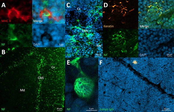
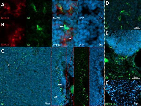
Recently, we found abundant innervation of antigen presenting cells that were reached and enclosed by single neurites. These neurally hard-wired antigen presenting cells (wAPC) could be observed in the T-cell zone of superficial cervical lymph nodes of rats and other mammalians, including humans.
As a consequence, we investigated lymph nodes at many different anatomical positions as well as all primary and secondary lymphoid organs (SLO) in rodents for a similar morphology of innervation regarding antigen presenting cells known in those tissues.
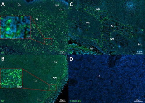
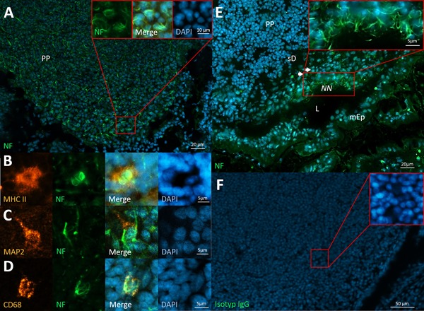
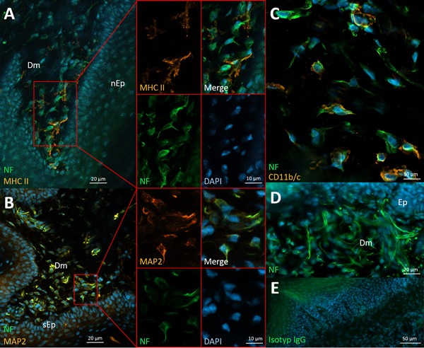
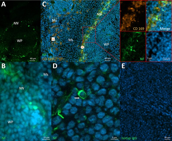
As a result, we confirmed wAPC in lymph nodes independent from their draining areas and anatomical positions but also in all other T-cell zones of lymphoid organs, like Peyer's patches, NALT and BALT, as well as in the thymic medulla. Other cells were innervated in a similar fashion but with seemingly missing antigen presenting capacity. Both types of innervated immune cells were observed as being also present in the dermis of the skin. Only in the spleen wAPC could not be detected. Beyond this systematic finding, we also found another regular phenomenon: a dense network of neurites that stained for neurofilament always in antigen entrance areas of lymphoid organs (subsinoidal layer of lymph nodes, subepithelial dome of Peyer's patches, subsinoidal layer of the splenic white pulp, margins of NALT and BALT). Lastly, also thymic epithelial cells (TEC) restricted to the corticomedullary junction of the thymus showed similar neurofilament staining.
Therefore, we propose much more hard-wired and probably afferent connections between lymphoid organs and the central nervous system than is hitherto known.
最近,我们发现大量的抗原呈递细胞被单一的神经突到达和包围,这些神经连接的抗原呈递细胞(wAPC)可以在大鼠和其他哺乳动物(包括人类)的浅表颈淋巴结的 T 细胞区观察到。
因此,我们研究了许多不同解剖位置的淋巴结以及所有啮齿动物的初级和次级淋巴器官(SLO),以寻找已知在这些组织中存在的类似神经支配的抗原呈递细胞形态。
我们在独立于引流区域和解剖位置的淋巴结中,以及在所有其他 T 细胞区的淋巴器官中,如派尔氏斑、NALT 和 BALT 以及胸腺髓质中,都证实了 wAPC 的存在。其他细胞也以类似的方式被神经支配,但似乎没有抗原呈递能力。这两种类型的被神经支配的免疫细胞也被观察到存在于皮肤的真皮中。只有在脾脏中没有检测到 wAPC。除了这种系统的发现,我们还发现了另一种常见现象:神经丝染色的密集神经突网络总是存在于淋巴器官的抗原进入区域(淋巴结的窦状层、派尔氏斑的上皮下穹窿、脾白髓的窦状层、NALT 和 BALT 的边缘)。最后,胸腺上皮细胞(TEC)也局限于胸腺的皮质髓质交界处,也表现出类似的神经丝染色。
因此,我们提出了比目前所知更多的淋巴器官和中枢神经系统之间的硬性和可能的传入连接。