Department of Neurology, School of Medicine, Epilepsy Center, Second Affiliated Hospital, Zhejiang University, Hangzhou, China.
Epilepsy Center, Cleveland Clinic, Cleveland, OH, USA.
Epilepsia. 2018 May;59(5):982-992. doi: 10.1111/epi.14064. Epub 2018 Apr 10.
Focal cortical dysplasia (FCD) is a major pathology in patients undergoing surgical resection to treat pharmacoresistant epilepsy. Magnetic resonance imaging (MRI) postprocessing methods may provide essential help for detection of FCD. In this study, we utilized surface-based MRI morphometry and machine learning for automated lesion detection in a mixed cohort of patients with FCD type II from 3 different epilepsy centers.
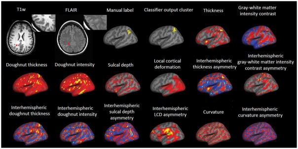
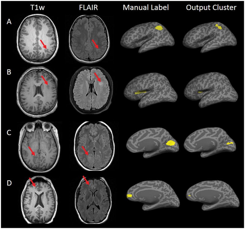
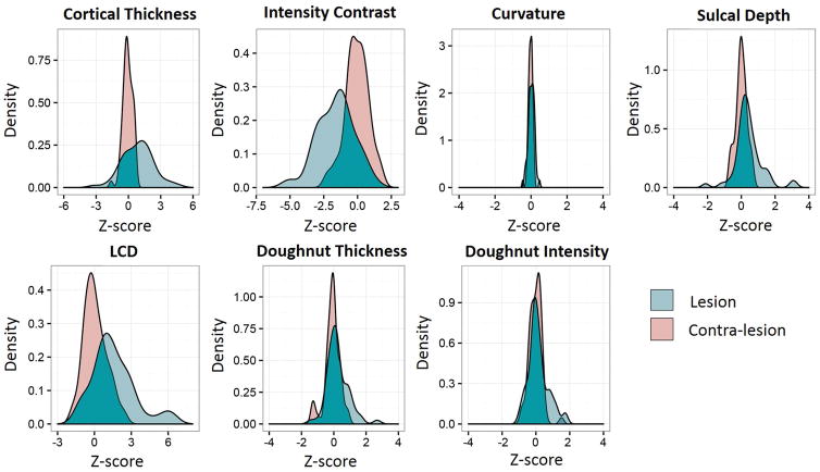
Sixty-one patients with pharmacoresistant epilepsy and histologically proven FCD type II were included in the study. The patients had been evaluated at 3 different epilepsy centers using 3 different MRI scanners. T1-volumetric sequence was used for postprocessing. A normal database was constructed with 120 healthy controls. We also included 35 healthy test controls and 15 disease test controls with histologically confirmed hippocampal sclerosis to assess specificity. Features were calculated and incorporated into a nonlinear neural network classifier, which was trained to identify lesional cluster. We optimized the threshold of the output probability map from the classifier by performing receiver operating characteristic (ROC) analyses. Success of detection was defined by overlap between the final cluster and the manual labeling. Performance was evaluated using k-fold cross-validation.
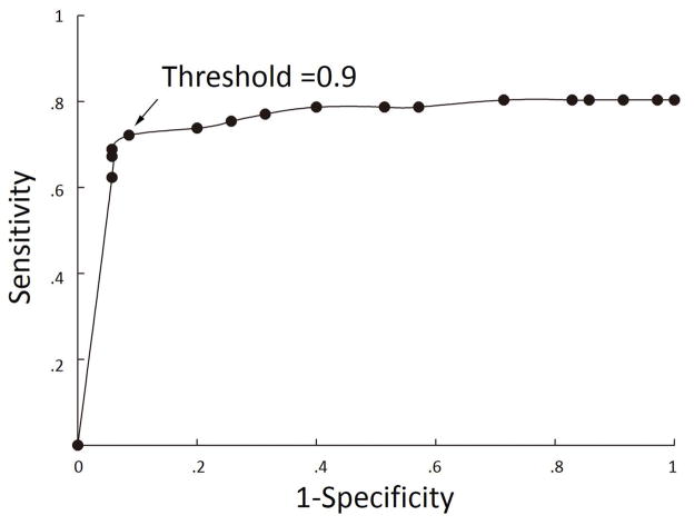
The threshold of 0.9 showed optimal sensitivity of 73.7% and specificity of 90.0%. The area under the curve for the ROC analysis was 0.75, which suggests a discriminative classifier. Sensitivity and specificity were not significantly different for patients from different centers, suggesting robustness of performance. Correct detection rate was significantly lower in patients with initially normal MRI than patients with unequivocally positive MRI. Subgroup analysis showed the size of the training group and normal control database impacted classifier performance.
Automated surface-based MRI morphometry equipped with machine learning showed robust performance across cohorts from different centers and scanners. The proposed method may be a valuable tool to improve FCD detection in presurgical evaluation for patients with pharmacoresistant epilepsy.
局灶性皮质发育不良(FCD)是接受手术切除以治疗药物难治性癫痫的患者的主要病理。磁共振成像(MRI)后处理方法可能为 FCD 的检测提供重要帮助。在这项研究中,我们利用基于表面的 MRI 形态计量学和机器学习,在来自 3 个不同癫痫中心的 FCD II 型混合队列中自动检测病变。
本研究纳入了 61 例药物难治性癫痫且组织学证实为 FCD II 型的患者。这些患者在 3 个不同的癫痫中心接受了 3 种不同的 MRI 扫描仪评估。使用 T1 容积序列进行后处理。使用 120 名健康对照构建正常数据库。我们还纳入了 35 名健康测试对照和 15 名组织学证实的海马硬化病测试对照,以评估特异性。计算特征并纳入非线性神经网络分类器,该分类器用于识别病变簇。我们通过执行接收器工作特征(ROC)分析来优化分类器输出概率图的阈值。通过最终簇与手动标记之间的重叠来定义检测的成功。使用 k 折交叉验证评估性能。
阈值为 0.9 时,灵敏度为 73.7%,特异性为 90.0%。ROC 分析的曲线下面积为 0.75,表明分类器具有区分能力。来自不同中心的患者的灵敏度和特异性没有显著差异,表明性能稳健。初始 MRI 正常的患者的正确检测率明显低于 MRI 阳性的患者。亚组分析表明,训练组的大小和正常对照数据库影响分类器性能。
配备机器学习的自动基于表面的 MRI 形态计量学在来自不同中心和扫描仪的队列中表现出稳健的性能。该方法可能是提高药物难治性癫痫患者术前评估中 FCD 检测的有价值工具。