Zuliani Carolina Coli, Bombini Mariana Freschi, Andrade Kleber Cursino de, Mamoni Ronei, Pereira Ana Helena, Coimbra Ibsen Bellini
Reumatologia, Clinica Medica, Universidade Estadual de Campinas, Campinas, SP, BR.
Tocoginecologia, Universidade Estadual de Campinas, Campinas, SP, BR.
Clinics (Sao Paulo). 2018;73:e268. doi: 10.6061/clinics/2018/e268. Epub 2018 Apr 5.
Articular cartilage is vulnerable to injuries and undergoes an irreversible degenerative process. The use of amniotic fluid mesenchymal stromal stem cells for the reconstruction of articular cartilage is a promising therapeutic alternative. The aim of this study was to investigate the chondrogenic potential of amniotic fluid mesenchymal stromal stem cells from human amniotic fluid from second trimester pregnant women in a micromass system (high-density cell culture) with TGF-β3 for 21 days.
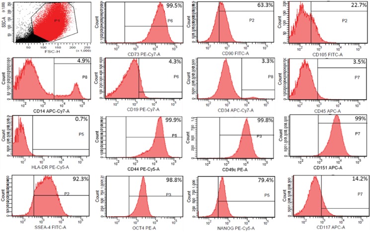
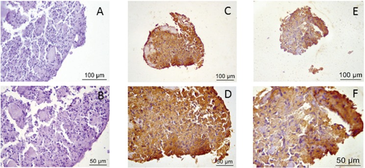
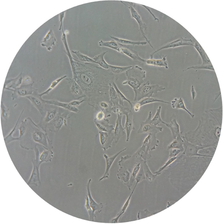
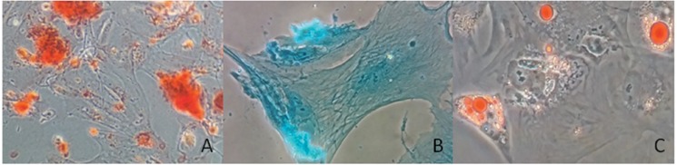
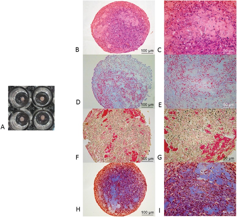
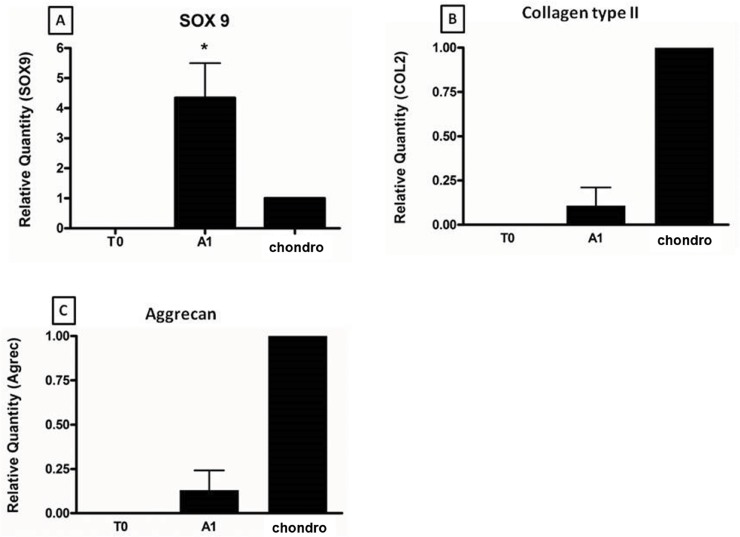
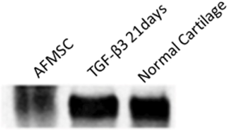
Micromass was performed using amniotic fluid mesenchymal stromal stem cells previously cultured in a monolayer. Chondrocytes from adult human normal cartilage were used as controls. After 21 days, chondrogenic potential was determined by measuring the expression of genes, such as SOX-9, type II collagen and aggrecan, in newly differentiated cells by real-time PCR (qRT-PCR). The production of type II collagen protein was observed by western blotting. Immunohistochemistry analysis was also performed to detect collagen type II and aggrecan. This study was approved by the local ethics committee.
SOX-9, aggrecan and type II collagen were expressed in newly differentiated chondrocytes. The expression of SOX-9 was significantly higher in newly differentiated chondrocytes than in adult cartilage. Collagen type II protein was also detected.
We demonstrate that stem cells from human amniotic fluid are a suitable source for chondrogenesis when cultured in a micromass system. amniotic fluid mesenchymal stromal stem cells are an extremely viable source for clinical applications, and our results suggest the possibility of using human amniotic fluid as a source of mesenchymal stem cells.
关节软骨易受损伤并经历不可逆的退变过程。使用羊水间充质基质干细胞重建关节软骨是一种有前景的治疗选择。本研究的目的是在微团培养系统(高密度细胞培养)中,用转化生长因子-β3处理来自孕中期孕妇羊水的间充质基质干细胞21天,以研究其软骨形成潜能。
使用先前单层培养的羊水间充质基质干细胞进行微团培养。将来自成人正常软骨的软骨细胞用作对照。21天后,通过实时聚合酶链反应(qRT-PCR)测量新分化细胞中SOX-9、II型胶原蛋白和聚集蛋白聚糖等基因的表达,以确定软骨形成潜能。通过蛋白质印迹法观察II型胶原蛋白的产生。还进行免疫组织化学分析以检测II型胶原蛋白和聚集蛋白聚糖。本研究获得当地伦理委员会的批准。
新分化的软骨细胞中表达了SOX-9、聚集蛋白聚糖和II型胶原蛋白。新分化的软骨细胞中SOX-9的表达明显高于成人软骨。还检测到了II型胶原蛋白。
我们证明,在微团培养系统中培养时,人羊水干细胞是软骨形成的合适来源。羊水间充质基质干细胞是临床应用中极具可行性的来源,我们的结果表明使用人羊水作为间充质干细胞来源的可能性。