Rheumatology Unit, Department of Clinical Medicine, School of Medical Sciences, State University of Campinas (UNICAMP), 126 Tessália Vieira de Camargo Street, Campinas, SP, CEP 13083-887, Brazil.
Department of Gynecology and Obstetrics, School of Medicine, State University of Campinas (UNICAMP), 101 Alexander Fleming Street, Campinas, SP, CEP 13083-891, Brazil.
Sci Rep. 2021 Feb 4;11(1):3063. doi: 10.1038/s41598-021-82341-x.
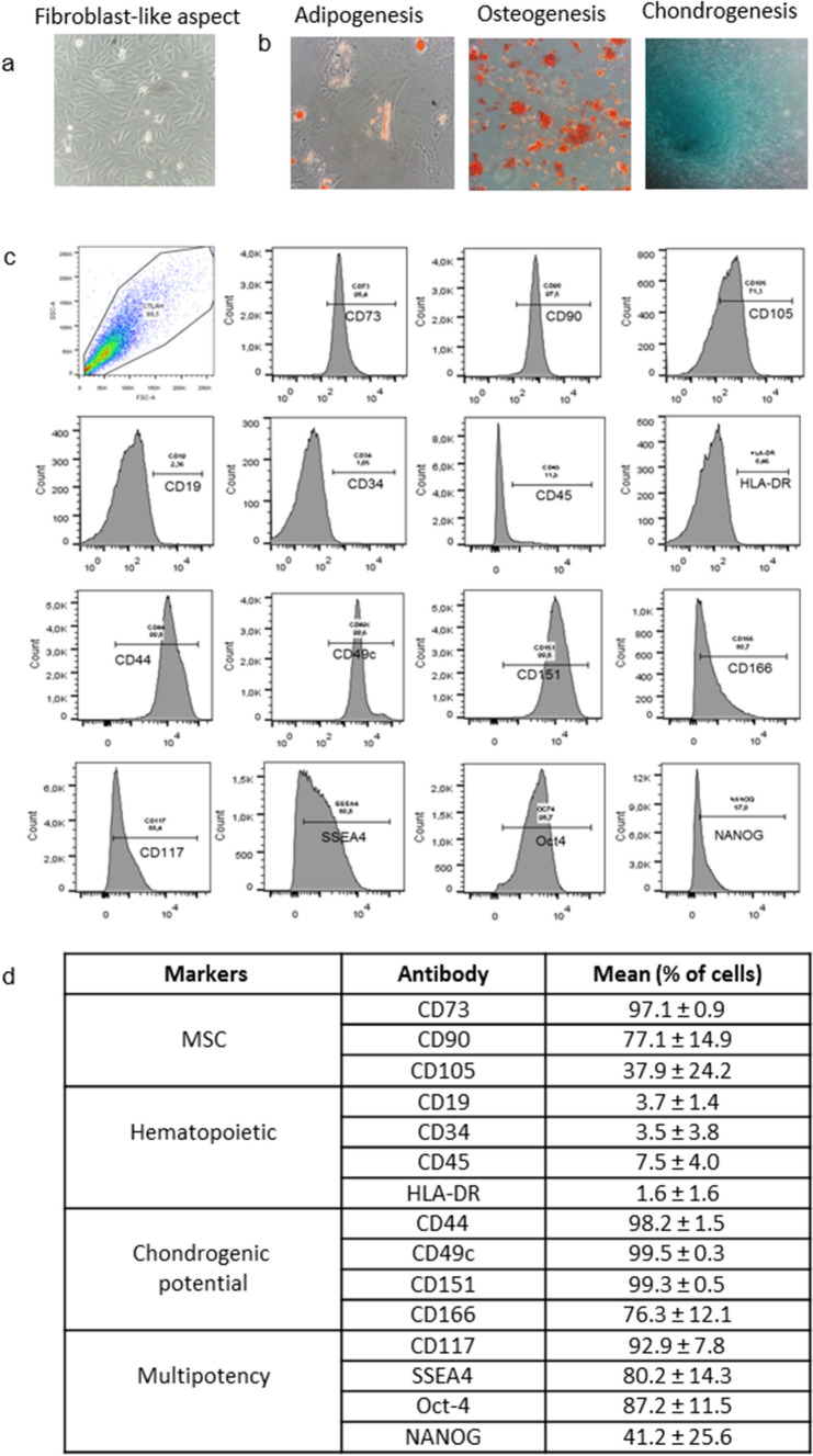
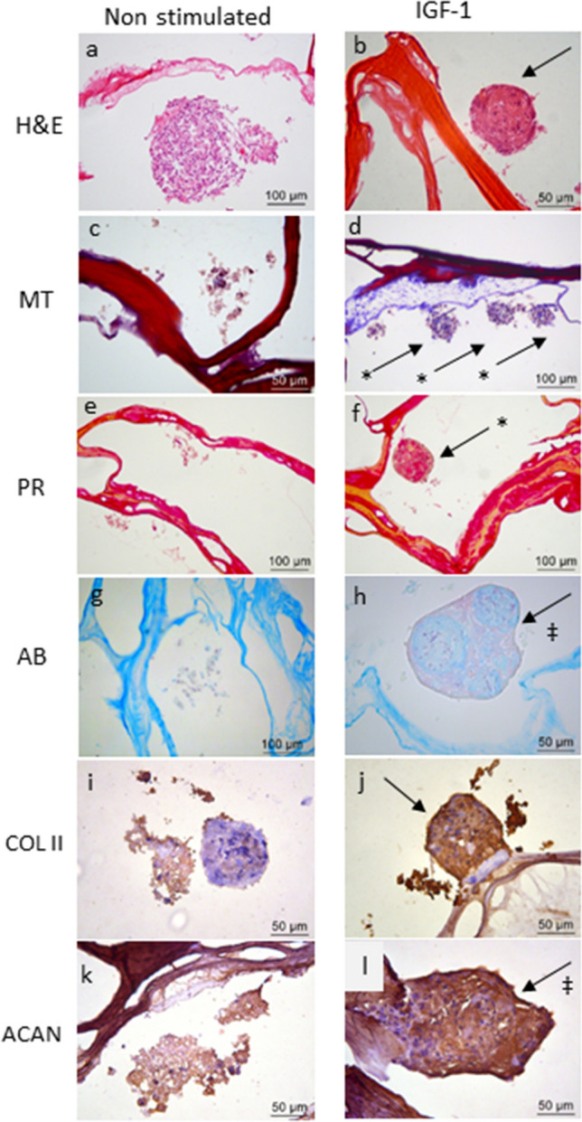
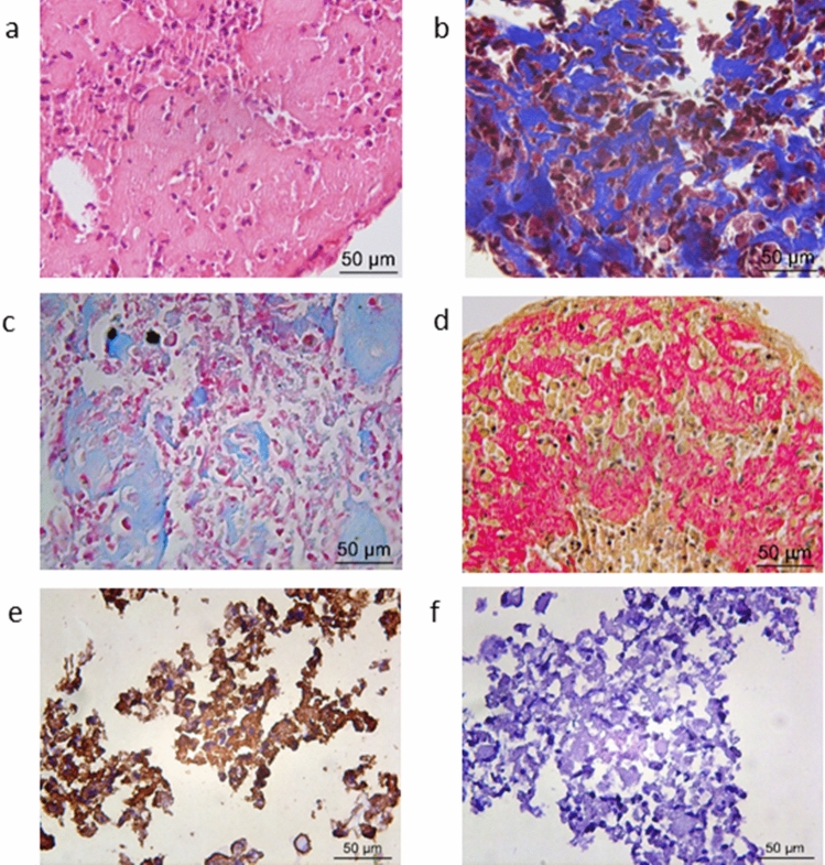
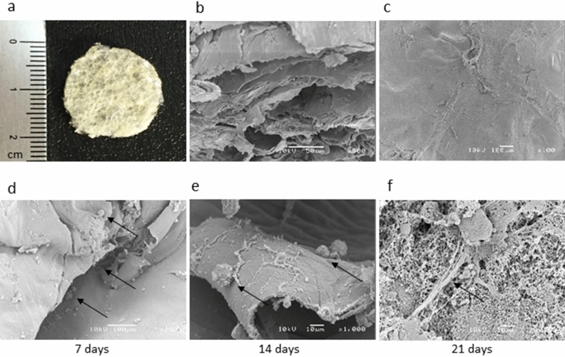
Articular chondral lesions, caused either by trauma or chronic cartilage diseases such as osteoarthritis, present very low ability to self-regenerate. Thus, their current management is basically symptomatic, progressing very often to invasive procedures or even arthroplasties. The use of amniotic fluid stem cells (AFSCs), due to their multipotentiality and plasticity, associated with scaffolds, is a promising alternative for the reconstruction of articular cartilage. Therefore, this study aimed to investigate the chondrogenic potential of AFSCs in a micromass system (high-density cell culture) under insulin-like growth factor 1 (IGF-1) stimuli, as well as to look at their potential to differentiate directly when cultured in a porous chitosan-xanthan (CX) scaffold. The experiments were performed with a CD117 positive cell population, with expression of markers (CD117, SSEA-4, Oct-4 and NANOG), selected from AFSCs, after immunomagnetic separation. The cells were cultured in both a micromass system and directly in the scaffold, in the presence of IGF-1. Differentiation to chondrocytes was confirmed by histology and by using immunohistochemistry. The construct cell-scaffold was also analyzed by scanning electron microscopy (SEM). The results demonstrated the chondrogenic potential of AFSCs cultivated directly in CX scaffolds and also in the micromass system. Such findings support and stimulate future studies using these constructs in osteoarthritic animal models.
关节软骨损伤可由创伤或慢性软骨疾病(如骨关节炎)引起,其自我再生能力非常低。因此,目前的治疗方法主要是对症治疗,常常会发展为侵袭性手术甚至关节置换术。由于羊水干细胞 (AFSCs) 具有多能性和可塑性,并与支架结合,因此是重建关节软骨的有前途的替代方法。因此,本研究旨在探讨在胰岛素样生长因子 1 (IGF-1) 刺激下,AFSCs 在微团系统(高密度细胞培养)中的软骨生成潜力,以及在多孔壳聚糖-黄原胶 (CX) 支架中直接培养时分化的潜力。通过免疫磁分离,从 AFSCs 中选择表达标记物(CD117、SSEA-4、Oct-4 和 NANOG)的 CD117 阳性细胞群体进行实验。将细胞分别在微团系统和支架中培养,在 IGF-1 的存在下进行分化。通过组织学和免疫组织化学证实向软骨细胞的分化。通过扫描电子显微镜 (SEM) 对细胞-支架构建体进行了分析。结果表明,AFSCs 直接在 CX 支架和微团系统中均具有软骨生成潜力。这些发现支持并激发了未来在骨关节炎动物模型中使用这些构建体的研究。