Mater Research Institute-University of Queensland, Woolloongabba, QLD 4102, Australia.
Northern Institute for Cancer Research, Newcastle University, Newcastle upon Tyne NE1 7RU, United Kingdom.
Genome Res. 2018 May;28(5):639-653. doi: 10.1101/gr.226993.117. Epub 2018 Apr 11.
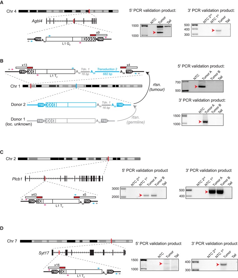
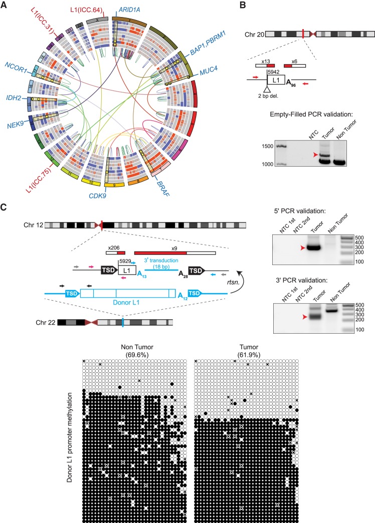
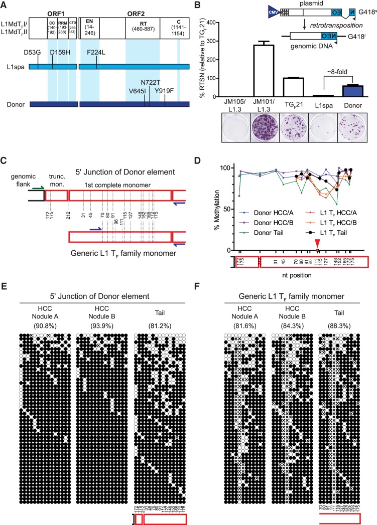
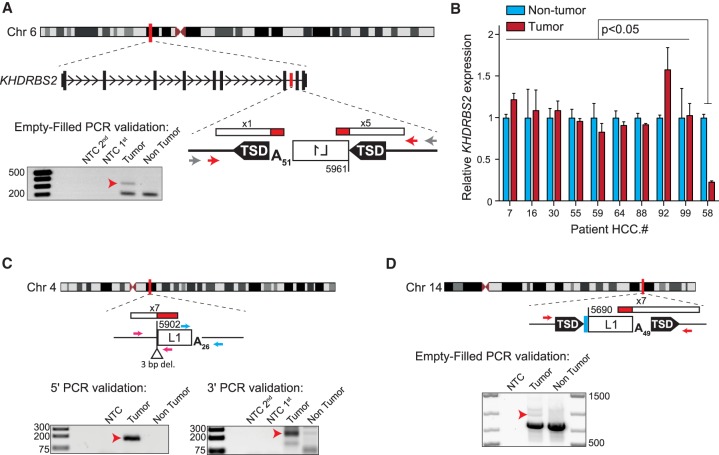
The retrotransposon Long Interspersed Element 1 (LINE-1 or L1) is a continuing source of germline and somatic mutagenesis in mammals. Deregulated L1 activity is a hallmark of cancer, and L1 mutagenesis has been described in numerous human malignancies. We previously employed retrotransposon capture sequencing (RC-seq) to analyze hepatocellular carcinoma (HCC) samples from patients infected with hepatitis B or hepatitis C virus and identified L1 variants responsible for activating oncogenic pathways. Here, we have applied RC-seq and whole-genome sequencing (WGS) to an mouse model of hepatic carcinogenesis and demonstrated for the first time that L1 mobilization occurs in murine tumors. In 12 HCC nodules obtained from 10 animals, we validated four somatic L1 insertions by PCR and capillary sequencing, including T subfamily elements, and one G subfamily example. One of the T insertions carried a 3' transduction, allowing us to identify its donor L1 and to demonstrate that this full-length T element retained retrotransposition capacity in cultured cancer cells. Using RC-seq, we also identified eight tumor-specific L1 insertions from 25 HCC patients with a history of alcohol abuse. Finally, we used RC-seq and WGS to identify three tumor-specific L1 insertions among 10 intra-hepatic cholangiocarcinoma (ICC) patients, including one insertion traced to a donor L1 on Chromosome 22 known to be highly active in other cancers. This study reveals L1 mobilization as a common feature of hepatocarcinogenesis in mammals, demonstrating that the phenomenon is not restricted to human viral HCC etiologies and is encountered in murine liver tumors.
长散布元件 1(LINE-1 或 L1)是哺乳动物种系和体细胞突变的持续来源。L1 活性失调是癌症的一个标志,并且已经在许多人类恶性肿瘤中描述了 L1 突变。我们之前采用逆转录转座子捕获测序(RC-seq)分析了乙型肝炎或丙型肝炎病毒感染患者的肝细胞癌(HCC)样本,并确定了导致致癌途径激活的 L1 变体。在这里,我们将 RC-seq 和全基因组测序(WGS)应用于肝致癌发生的小鼠模型,并首次证明 L1 动员发生在鼠肿瘤中。在从 10 只动物获得的 12 个 HCC 结节中,我们通过 PCR 和毛细管测序验证了四个体细胞 L1 插入,包括 T 亚家族元件和一个 G 亚家族实例。一个 T 插入带有 3'转导,使我们能够识别其供体 L1,并证明这个全长 T 元素在培养的癌细胞中保留了逆转录转位能力。使用 RC-seq,我们还从 25 名有酗酒史的 HCC 患者中鉴定了 8 个肿瘤特异性 L1 插入。最后,我们使用 RC-seq 和 WGS 在 10 名肝内胆管癌(ICC)患者中鉴定了三个肿瘤特异性 L1 插入,包括一个插入可以追溯到已知在其他癌症中高度活跃的染色体 22 上的供体 L1。这项研究揭示了 L1 动员作为哺乳动物肝癌发生的一个共同特征,表明这种现象不仅限于人类病毒性 HCC 病因,并且在鼠肝肿瘤中也有发现。