Center for Biomedical Informatics, Wake Forest School of Medicine, Winston Salem, NC, United States of America.
Department of Biomedical Informatics, The Ohio State University, Columbus, OH, United States of America.
PLoS One. 2018 Apr 12;13(4):e0195621. doi: 10.1371/journal.pone.0195621. eCollection 2018.
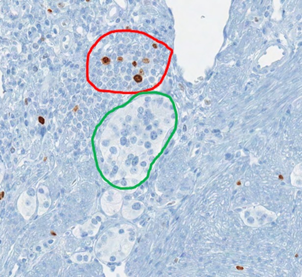
The World Health Organization (WHO) has clear guidelines regarding the use of Ki67 index in defining the proliferative rate and assigning grade for pancreatic neuroendocrine tumor (NET). WHO mandates the quantification of Ki67 index by counting at least 500 positive tumor cells in a hotspot. Unfortunately, Ki67 antibody may stain both tumor and non-tumor cells as positive depending on the phase of the cell cycle. Likewise, the counter stain labels both tumor and non-tumor as negative. This non-specific nature of Ki67 stain and counter stain therefore hinders the exact quantification of Ki67 index. To address this problem, we present a deep learning method to automatically differentiate between NET and non-tumor regions based on images of Ki67 stained biopsies. Transfer learning was employed to recognize and apply relevant knowledge from previous learning experiences to differentiate between tumor and non-tumor regions. Transfer learning exploits a rich set of features previously used to successfully categorize non-pathology data into 1,000 classes. The method was trained and validated on a set of whole-slide images including 33 NETs subject to Ki67 immunohistochemical staining using a leave-one-out cross-validation. When applied to 30 high power fields (HPF) and assessed against a gold standard (evaluation by two expert pathologists), the method resulted in a high sensitivity of 97.8% and specificity of 88.8%. The deep learning method developed has the potential to reduce pathologists' workload by directly identifying tumor boundaries on images of Ki67 stained slides. Moreover, it has the potential to replace sophisticated and expensive imaging methods which are recently developed for identification of tumor boundaries in images of Ki67-stained NETs.
世界卫生组织 (WHO) 对 Ki67 指数在定义胰腺神经内分泌肿瘤 (NET) 的增殖率和分级中的使用有明确的指导方针。WHO 要求通过在热点区域计数至少 500 个阳性肿瘤细胞来量化 Ki67 指数。不幸的是,Ki67 抗体可能根据细胞周期的阶段将肿瘤细胞和非肿瘤细胞都染色为阳性。同样,计数器染色将肿瘤和非肿瘤都标记为阴性。因此,Ki67 染色和计数器染色的这种非特异性阻碍了 Ki67 指数的准确量化。为了解决这个问题,我们提出了一种基于 Ki67 染色活检图像的深度学习方法,自动区分 NET 和非肿瘤区域。迁移学习用于识别和应用以前学习经验中的相关知识,以区分肿瘤和非肿瘤区域。迁移学习利用了一组丰富的特征,这些特征以前曾被成功地用于将非病理学数据分类为 1000 个类别。该方法在一组全幻灯片图像上进行训练和验证,包括 33 个接受 Ki67 免疫组织化学染色的 NET,并使用留一法交叉验证。当应用于 30 个高倍视野 (HPF) 并与金标准(由两名专家病理学家评估)进行评估时,该方法的灵敏度为 97.8%,特异性为 88.8%。开发的深度学习方法有可能通过直接识别 Ki67 染色幻灯片上的肿瘤边界来减轻病理学家的工作量。此外,它还有可能取代最近为识别 Ki67 染色 NET 图像中的肿瘤边界而开发的复杂且昂贵的成像方法。