European Synchrotron Radiation Facility, BP 220, 38043 Grenoble, France.
European Molecular Biology Laboratory, Hamburg Outstation, Notkestrasse 85, 22607 Hamburg, Germany.
Acta Crystallogr D Struct Biol. 2018 Apr 1;74(Pt 4):355-365. doi: 10.1107/S2059798318002735. Epub 2018 Apr 6.
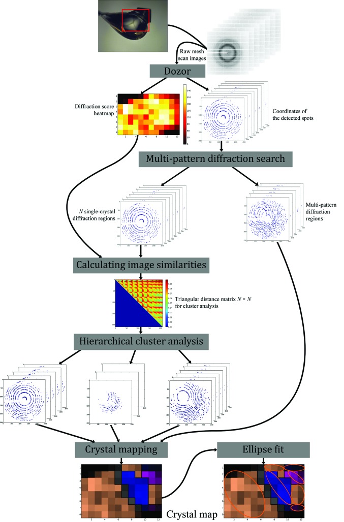
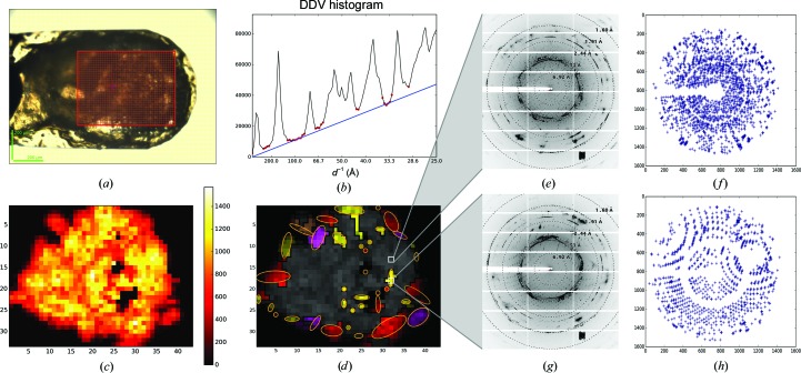
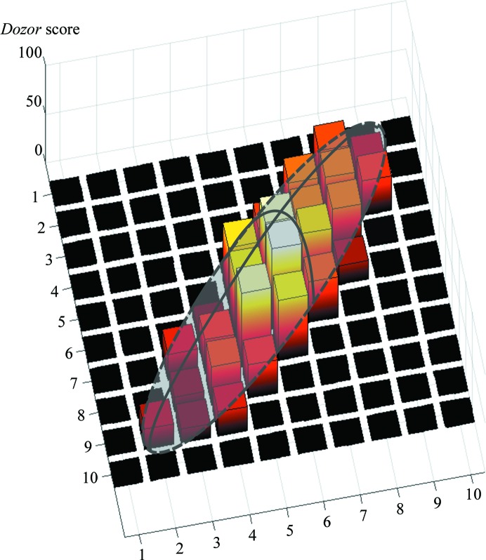
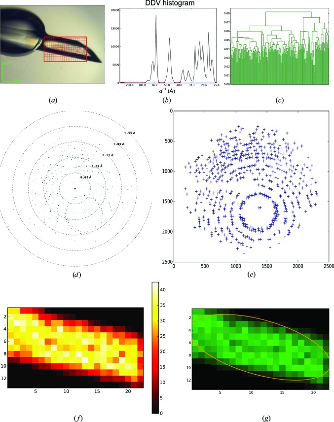
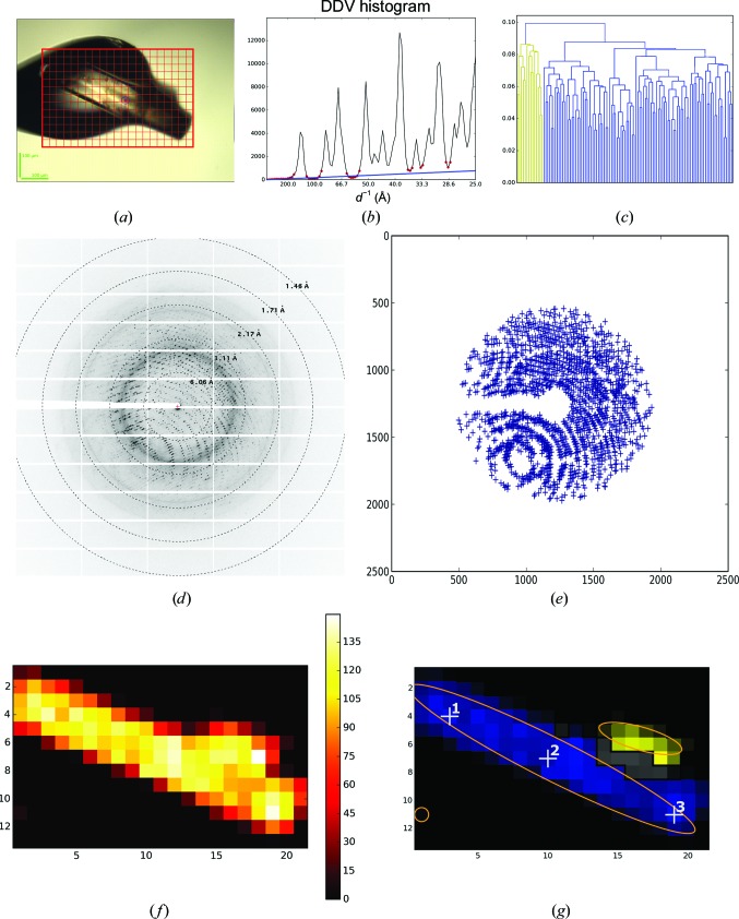
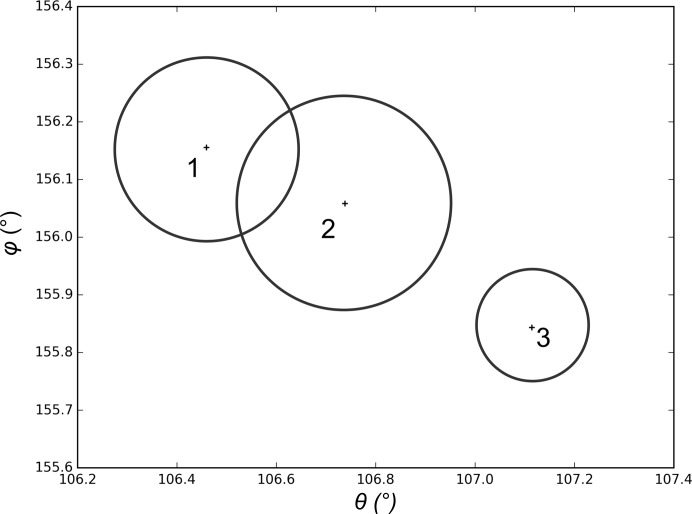
In macromolecular crystallography, mesh (raster) scans are carried out either as part of X-ray-based crystal-centring routines or to identify positions on the sample holder from which diffraction images can be collected. Here, the methods used in MeshBest, software which automatically analyses diffraction images collected during a mesh scan and produces a two-dimensional crystal map showing estimates of the dimensions, centre positions and diffraction qualities of each crystal contained in the mesh area, are presented. Sample regions producing diffraction images resulting from the superposition of more than one crystal are also distinguished from regions with single-crystal diffraction. The applicability of the method is demonstrated using several cases.
在大分子晶体学中,网格(光栅)扫描是作为基于 X 射线的晶体定位程序的一部分进行的,或者是为了确定可以收集衍射图像的样品架上的位置。本文介绍了 MeshBest 软件中使用的方法,该软件可自动分析网格扫描过程中收集的衍射图像,并生成二维晶体图,显示网格区域内每个晶体的尺寸、中心位置和衍射质量的估计值。软件还可以区分产生多个晶体叠加衍射图像的区域和具有单晶衍射的区域。该方法的适用性通过几个案例得到了证明。