Department of Medical, Oral and Biotechnological Sciences, University "G. d'Annunzio", Chieti, Italy.
IRCCS Centro Neurolesi "Bonino Pulejo", Messina, Italy.
Stem Cell Res Ther. 2018 Apr 13;9(1):104. doi: 10.1186/s13287-018-0850-0.
The role of bone tissue engineering in the field of regenerative medicine has been a main research topic over the past few years. There has been much interest in the use of three-dimensional (3D) engineered scaffolds (PLA) complexed with human gingival mesenchymal stem cells (hGMSCs) as a new therapeutic strategy to improve bone tissue regeneration. These devices can mimic a more favorable endogenous microenvironment for cells in vivo by providing 3D substrates which are able to support cell survival, proliferation and differentiation. The present study evaluated the in vitro and in vivo capability of bone defect regeneration of 3D PLA, hGMSCs, extracellular vesicles (EVs), or polyethyleneimine (PEI)-engineered EVs (PEI-EVs) in the following experimental groups: 3D-PLA, 3D-PLA + hGMSCs, 3D-PLA + EVs, 3D-PLA + EVs + hGMSCs, 3D-PLA + PEI-EVs, 3D-PLA + PEI-EVs + hGMSCs.
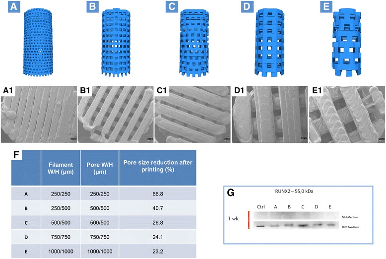
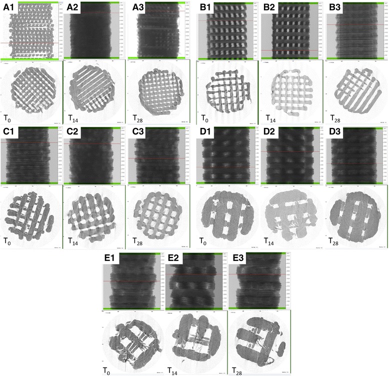
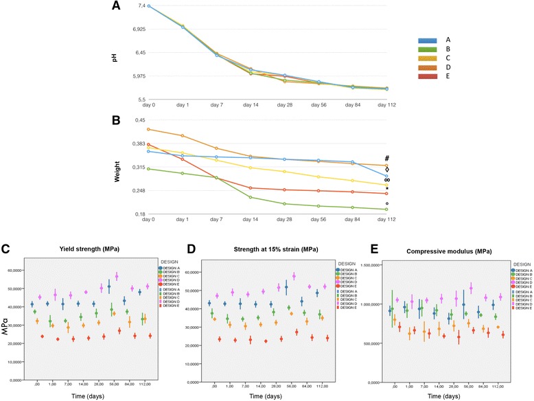
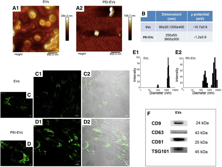
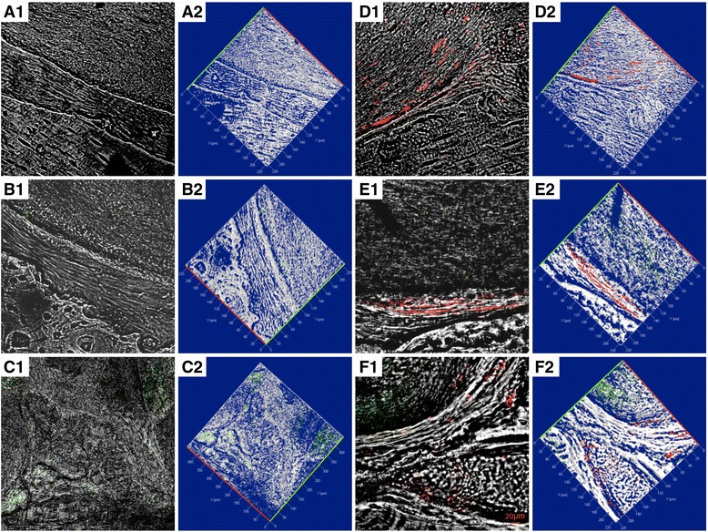
The structural parameters of the scaffold were evaluated using both scanning electron microscopy and nondestructive microcomputed tomography. Nanotopographic surface features were investigated by means of atomic force microscopy. Scaffolds showed a statistically significant mass loss along the 112-day evaluation.
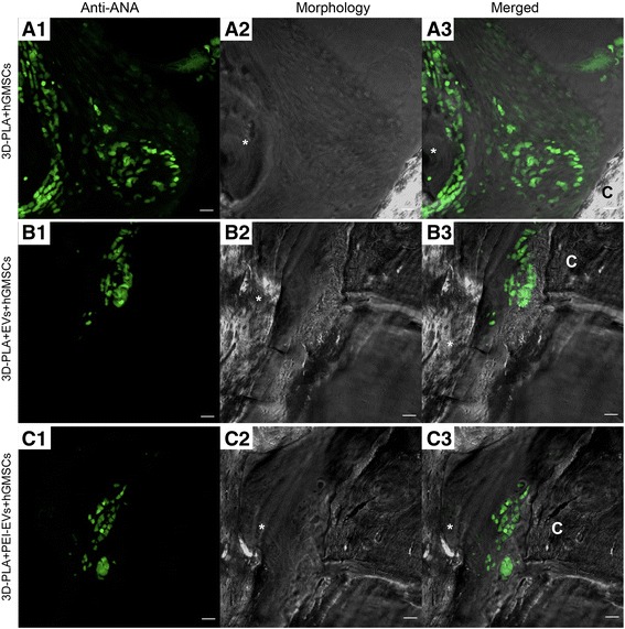
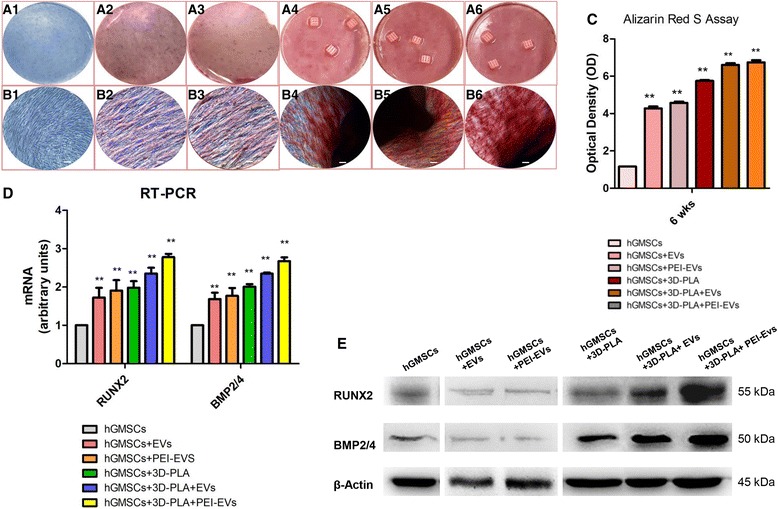
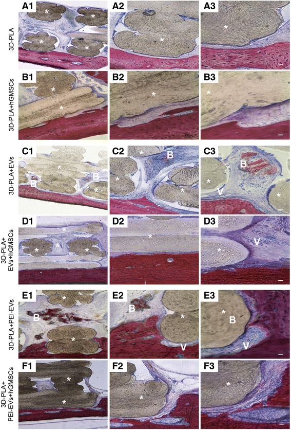
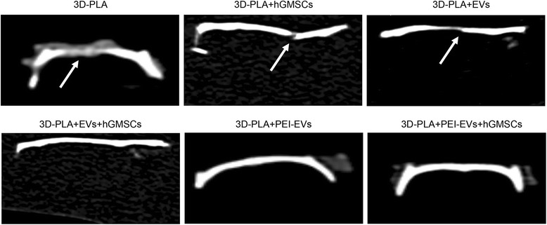
Our in vitro results revealed that both 3D-PLA + EVs + hGMSCs and 3D-PLA + PEI-EVs + hGMSCs showed no cytotoxicity. However, 3D-PLA + PEI-EVs + hGMSCs exhibited greater osteogenic inductivity as revealed by morphological evaluation and transcriptomic analysis performed by next-generation sequencing (NGS). In addition, in vivo results showed that 3D-PLA + PEI-EVs + hGMSCs and 3D-PLA + PEI-EVs scaffolds implanted in rats subjected to cortical calvaria bone tissue damage were able to improve bone healing by showing better osteogenic properties. These results were supported also by computed tomography evaluation that revealed the repair of bone calvaria damage.
The re-establishing of the integrity of the bone lesions could be a promising strategy in the treatment of accidental or surgery trauma, especially for cranial bones.
在再生医学领域,骨组织工程一直是一个主要的研究课题。人们对使用三维(3D)工程支架(PLA)与人类牙龈间充质干细胞(hGMSCs)复合作为一种新的治疗策略来改善骨组织再生非常感兴趣。这些装置可以通过提供能够支持细胞存活、增殖和分化的 3D 基质,模拟更有利于细胞在体内生存的内源性微环境。本研究评估了以下实验组中 3D PLA、hGMSCs、细胞外囊泡(EVs)或聚乙烯亚胺(PEI)工程 EVs(PEI-EVs)的体外和体内骨缺损再生能力:3D-PLA、3D-PLA+hGMSCs、3D-PLA+EVs、3D-PLA+EVs+hGMSCs、3D-PLA+PEI-EVs、3D-PLA+PEI-EVs+hGMSCs。
使用扫描电子显微镜和无损微计算机断层扫描评估支架的结构参数。原子力显微镜用于研究纳米拓扑表面特征。支架在 112 天的评估过程中表现出统计学上显著的质量损失。
我们的体外结果表明,3D-PLA+EVs+hGMSCs 和 3D-PLA+PEI-EVs+hGMSCs 均无细胞毒性。然而,3D-PLA+PEI-EVs+hGMSCs 通过形态学评估和下一代测序(NGS)进行的转录组分析显示出更强的成骨诱导性。此外,体内结果表明,在大鼠皮质颅骨骨组织损伤模型中植入 3D-PLA+PEI-EVs+hGMSCs 和 3D-PLA+PEI-EVs 支架能够通过表现出更好的成骨特性来改善骨愈合。这些结果也得到了计算机断层扫描评估的支持,该评估显示了颅骨损伤的修复。
重建骨病变的完整性可能是治疗意外或手术创伤的一种有前途的策略,特别是对于颅骨。