• 文献检索
  • 文档翻译
  • 深度研究
  • 学术资讯
  • Suppr Zotero 插件Zotero 插件
  • 邀请有礼
  • 套餐&价格
  • 历史记录
应用&插件
Suppr Zotero 插件Zotero 插件浏览器插件Mac 客户端Windows 客户端微信小程序
定价
高级版会员购买积分包购买API积分包
服务
文献检索文档翻译深度研究API 文档MCP 服务
关于我们
关于 Suppr公司介绍联系我们用户协议隐私条款
关注我们

Suppr 超能文献

核心技术专利:CN118964589B侵权必究
粤ICP备2023148730 号-1Suppr @ 2026

文献检索

告别复杂PubMed语法,用中文像聊天一样搜索,搜遍4000万医学文献。AI智能推荐,让科研检索更轻松。

立即免费搜索

文件翻译

保留排版,准确专业,支持PDF/Word/PPT等文件格式,支持 12+语言互译。

免费翻译文档

深度研究

AI帮你快速写综述,25分钟生成高质量综述,智能提取关键信息,辅助科研写作。

立即免费体验

用于乳腺病变鉴别诊断的实时弹性成像

Real-Time Elastography for Differentiation of Breast Lesions.

作者信息

Yılmaz Ebru, Yılmaz Ayhan, Aslan Ahmet, Inan Ibrahim, Evren Mujgan Calıskan, Tekesin Kemal

机构信息

Department of Radiology, Gaziosmanpaşa Taksim Training and Research Hospital, Istanbul, Turkey.

Department of Radiology, Bezmialem Vakif University Hospital, Istanbul, Turkey.

出版信息

Pol J Radiol. 2017 Nov 17;82:664-669. doi: 10.12659/PJR.902596. eCollection 2017.

DOI:10.12659/PJR.902596
PMID:29657632
原文链接:https://pmc.ncbi.nlm.nih.gov/articles/PMC5894028/
Abstract

BACKROUNG

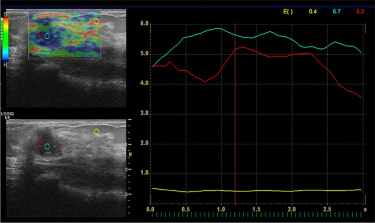
To investigate the diagnostic performance of the elastography-based strain index ratio in the differential diagnosis of malignant and benign breast lesions.

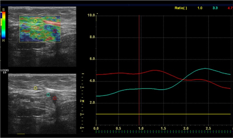
MATERIAL/METHODS: Seventy-nine breast masses that were classified as BI-RADS category 3, 4, and 5 on B-mode ultrasonography (US) were further prospectively evaluated by real-time sonoelastography (RTE). To obtain an optimal cut-off value of the strain ratio for differentiating between malignant and benign breast lesions, RTE findings were compared with histopathology of core needle biopsy samples or with ultrasound follow-up data of the analyzed masses.

RESULTS

Seventy-nine breast lesions [BI-RADS category 3 (n=15), BI-RADS category 4 (n=34), and BI-RADS category 5 (n=30)] were classified as malignant (n=36) or benign (n=43). The mean strain index value was 6.59±3.44 (range 0.6-14) for malignant lesions and 2.79±2.16 (range 0.6-8.7) for benign lesions, respectively (p<0.05). As regards the detection of malignant lesions, US was characterized by sensitivity and specificity of 100% (CI 95%; 88-100) and 90% (CI 95%; 76-97), respectively. When an optimal value of the strain ratio (4.25) was obtained by ROC curve analysis, the sensitivity and specificity for diagnosing malignant lesions were 86% (CI 95%; 70-95) and 76% (CI 95%; 60-87), respectively.

CONCLUSIONS

RTE can play an important role in the differentiation between malignant and benign breast masses, but it should be used in conjunction with ultrasonography.

摘要

背景

探讨基于弹性成像的应变指数比在乳腺良恶性病变鉴别诊断中的诊断性能。

材料/方法:对79例在B型超声(US)检查中被分类为BI-RADS 3、4和5类的乳腺肿块进行实时超声弹性成像(RTE)前瞻性评估。为获得用于区分乳腺良恶性病变的应变比最佳临界值,将RTE检查结果与粗针活检样本的组织病理学结果或所分析肿块的超声随访数据进行比较。

结果

79例乳腺病变[BI-RADS 3类(n = 15)、BI-RADS 4类(n = 34)和BI-RADS 5类(n = 30)]被分类为恶性(n = 36)或良性(n = 43)。恶性病变的平均应变指数值分别为6.59±3.44(范围0.6 - 14),良性病变为2.79±2.16(范围0.6 - 8.7)(p<0.05)。在恶性病变检测方面,US的敏感性和特异性分别为100%(CI 95%;88 - 100)和90%(CI 95%;76 - 97)。通过ROC曲线分析获得应变比的最佳值(4.25)时,诊断恶性病变的敏感性和特异性分别为86%(CI 95%;70 - 95)和76%(CI 95%;60 - 87)。

结论

RTE在乳腺良恶性肿块的鉴别中可发挥重要作用,但应与超声联合使用。

https://cdn.ncbi.nlm.nih.gov/pmc/blobs/e93a/5894028/2301c7d19896/PJR-82-902596-g002.jpg
https://cdn.ncbi.nlm.nih.gov/pmc/blobs/e93a/5894028/8e4a3610d603/PJR-82-902596-g001.jpg
https://cdn.ncbi.nlm.nih.gov/pmc/blobs/e93a/5894028/2301c7d19896/PJR-82-902596-g002.jpg
https://cdn.ncbi.nlm.nih.gov/pmc/blobs/e93a/5894028/8e4a3610d603/PJR-82-902596-g001.jpg
https://cdn.ncbi.nlm.nih.gov/pmc/blobs/e93a/5894028/2301c7d19896/PJR-82-902596-g002.jpg

相似文献

1
Real-Time Elastography for Differentiation of Breast Lesions.用于乳腺病变鉴别诊断的实时弹性成像
Pol J Radiol. 2017 Nov 17;82:664-669. doi: 10.12659/PJR.902596. eCollection 2017.
2
Diagnostic performance of ultrasound strain elastography for differentiation of malignant breast lesions.超声应变弹性成像对乳腺恶性病变的诊断效能
Clin Hemorheol Microcirc. 2019;71(2):237-247. doi: 10.3233/CH-189415.
3
Can strain elastography combined with ultrasound breast imaging reporting and data system be a more effective method in the differentiation of benign and malignant breast lesions?应变弹性成像结合乳腺超声影像报告和数据系统能否成为鉴别乳腺良恶性病变的更有效方法?
J Med Ultrason (2001). 2017 Oct;44(4):289-296. doi: 10.1007/s10396-017-0772-y. Epub 2017 Feb 2.
4
A set of shear wave elastography quantitative parameters combined with ultrasound BI-RADS to assess benign and malignant breast lesions.一组剪切波弹性成像定量参数联合超声BI-RADS用于评估乳腺良恶性病变。
Ultrasound Med Biol. 2015 Apr;41(4):960-6. doi: 10.1016/j.ultrasmedbio.2014.11.014. Epub 2015 Feb 17.
5
What Help Could Ultrasound Elastography Give to the Diagnosis of Breast Papillary Lesions?超声弹性成像对乳腺乳头状病变的诊断有何帮助?
Ultrasound Med Biol. 2017 May;43(5):903-910. doi: 10.1016/j.ultrasmedbio.2017.01.004. Epub 2017 Feb 27.
6
Comparison and Combination of Strain and Shear Wave Elastography of Breast Masses for Differentiation of Benign and Malignant Lesions by Quantitative Assessment: Preliminary Study.通过定量评估比较和联合应用乳腺肿块的应变弹性成像与剪切波弹性成像以鉴别良恶性病变:初步研究
J Ultrasound Med. 2018 Jan;37(1):99-109. doi: 10.1002/jum.14309. Epub 2017 Jul 8.
7
The Utility of the Fifth Edition of the BI-RADS Ultrasound Lexicon in Category 4 Breast Lesions: A Prospective Multicenter Study in China.第五版 BI-RADS 超声词汇在 4 类乳腺病变中的应用:中国前瞻性多中心研究。
Acad Radiol. 2022 Jan;29 Suppl 1:S26-S34. doi: 10.1016/j.acra.2020.06.027. Epub 2020 Aug 4.
8
Usefulness of ultrasound elastography in reducing the number of Breast Imaging Reporting and Data System category 3 lesions on ultrasonography.超声弹性成像在减少超声 BI-RADS 3 类病变数量中的作用。
Ultrasonography. 2014 Apr;33(2):98-104. doi: 10.14366/usg.13024. Epub 2014 Feb 26.
9
Diagnostic performance of quantitative shear wave elastography in the evaluation of solid breast masses: determination of the most discriminatory parameter.定量剪切波弹性成像在评估实性乳腺肿块中的诊断性能:最具鉴别力参数的确定。
AJR Am J Roentgenol. 2014 Sep;203(3):W328-36. doi: 10.2214/AJR.13.11693.
10
Multicenter study of ultrasound real-time tissue elastography in 779 cases for the assessment of breast lesions: improved diagnostic performance by combining the BI-RADS®-US classification system with sonoelastography.多中心研究 779 例超声实时组织弹性成像用于评估乳腺病变:结合 BI-RADS®-US 分类系统与声弹性成像提高诊断性能。
Ultraschall Med. 2010 Oct;31(5):484-91. doi: 10.1055/s-0029-1245282. Epub 2010 Apr 20.

引用本文的文献

1
Breast cancer imaging-clinical experience with two-dimensional-shear wave elastography: A retrospective study.乳腺癌成像——二维剪切波弹性成像的临床经验:一项回顾性研究。
World J Radiol. 2024 Oct 28;16(10):528-536. doi: 10.4329/wjr.v16.i10.528.
2
Strain Elastography Fat-to-Lesion Index Is Associated with Mammography BI-RADS Grading, Biopsy, and Molecular Phenotype in Breast Cancer.应变弹性成像脂肪至病变指数与乳腺癌的钼靶 BI-RADS 分级、活检和分子表型相关。
Biosensors (Basel). 2024 Feb 10;14(2):94. doi: 10.3390/bios14020094.
3
Determining the elastography strain ratio cut off value for differentiating benign from malignant breast lesions: systematic review and meta-analysis.

本文引用的文献

1
Comparison of shear-wave and strain ultrasound elastography in the differentiation of benign and malignant breast lesions.剪切波和应变成像超声弹性成像在鉴别良恶性乳腺病变中的比较。
AJR Am J Roentgenol. 2013 Aug;201(2):W347-56. doi: 10.2214/AJR.12.10416.
2
Clinical application of breast elastography: state of the art.乳腺弹性成像的临床应用:最新进展。
Eur J Radiol. 2014 Mar;83(3):429-37. doi: 10.1016/j.ejrad.2013.05.007. Epub 2013 Jun 18.
3
Diagnosis of solid breast lesions by elastography 5-point score and strain ratio method.
确定弹性成像应变比截断值以区分良恶性乳腺病变:系统评价和荟萃分析。
Cancer Imaging. 2022 Feb 12;22(1):12. doi: 10.1186/s40644-022-00447-5.
4
Graph-based rotational nonuniformity correction for localized compliance measurement in the human nasopharynx.基于图形的旋转非均匀性校正,用于人体鼻咽部局部顺应性测量
Biomed Opt Express. 2021 Mar 30;12(4):2508-2518. doi: 10.1364/BOE.419997. eCollection 2021 Apr 1.
5
Can strain elastography be used in reclassification of indeterminate breast lesions in BIRADS lexicon?: A prospective study.应变弹性成像能否用于重新分类乳腺影像报告和数据系统(BIRADS)词典中的不确定乳腺病变?一项前瞻性研究。
Indian J Radiol Imaging. 2020 Oct-Dec;30(4):493-499. doi: 10.4103/ijri.IJRI_425_19. Epub 2021 Jan 13.
6
Added value of strain elastography in the characterisation of breast lesions: A prospective study.应变弹性成像在乳腺病变特征描述中的附加价值:一项前瞻性研究。
Ultrasound. 2020 Aug;28(3):164-173. doi: 10.1177/1742271X20912762. Epub 2020 Mar 16.
弹性成像 5 分评分法和应变率比值法诊断实性乳腺病变。
Eur J Radiol. 2012 Nov;81(11):3245-9. doi: 10.1016/j.ejrad.2012.06.004. Epub 2012 Jun 27.
4
Screening US in patients with mammographically dense breasts: initial experience with Connecticut Public Act 09-41.乳腺钼靶摄影致密型乳腺患者的筛查:康涅狄格州公法 09-41 的初步经验。
Radiology. 2012 Oct;265(1):59-69. doi: 10.1148/radiol.12120621. Epub 2012 Jun 21.
5
Sonographic breast elastography: a primer.超声弹性成像在乳腺中的应用:入门篇。
J Ultrasound Med. 2012 May;31(5):773-83. doi: 10.7863/jum.2012.31.5.773.
6
Shear wave elastography for breast masses is highly reproducible.剪切波弹性成像技术在乳腺肿块中的重复性较高。
Eur Radiol. 2012 May;22(5):1023-32. doi: 10.1007/s00330-011-2340-y. Epub 2011 Dec 31.
7
Distinguishing benign from malignant masses at breast US: combined US elastography and color doppler US--influence on radiologist accuracy.乳腺超声鉴别良恶性肿块:超声弹性成像联合彩色多普勒超声——对放射科医生准确率的影响。
Radiology. 2012 Jan;262(1):80-90. doi: 10.1148/radiol.11110886. Epub 2011 Nov 14.
8
Qualitative and semiquantitative evaluations of solid breast lesions by sonoelastography.超声弹性成像对乳腺实性病变的定性和半定量评估
J Ultrasound Med. 2011 Feb;30(2):179-86. doi: 10.7863/jum.2011.30.2.179.
9
Using real-time tissue elastography for breast lesion evaluation: our initial experience.应用实时组织弹性成像技术评估乳腺病变:我们的初步经验。
J Ultrasound Med. 2010 Apr;29(4):551-63. doi: 10.7863/jum.2010.29.4.551.
10
Role of sonoelastography in characterising breast nodules. Preliminary experience with 120 lesions.超声弹性成像在乳腺结节特征描述中的作用。120 个病灶的初步经验。
Radiol Med. 2010 Jun;115(4):551-62. doi: 10.1007/s11547-010-0518-z. Epub 2010 Feb 22.