Department of Biomedical Sciences, College of Veterinary Medicine, Iowa State University, Ames, Iowa, United States of America.
Department of Pathobiological Sciences, School of Veterinary Medicine, University of Wisconsin-Madison, Madison, Wisconsin, United States of America.
PLoS Negl Trop Dis. 2018 Apr 16;12(4):e0006438. doi: 10.1371/journal.pntd.0006438. eCollection 2018 Apr.
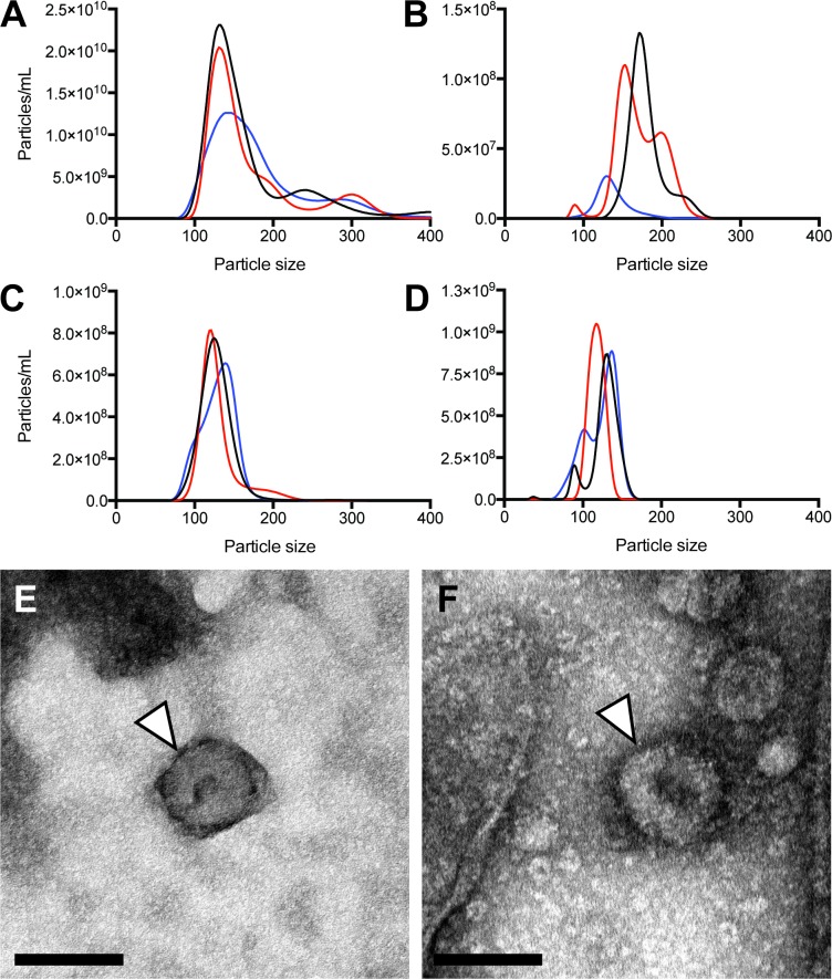
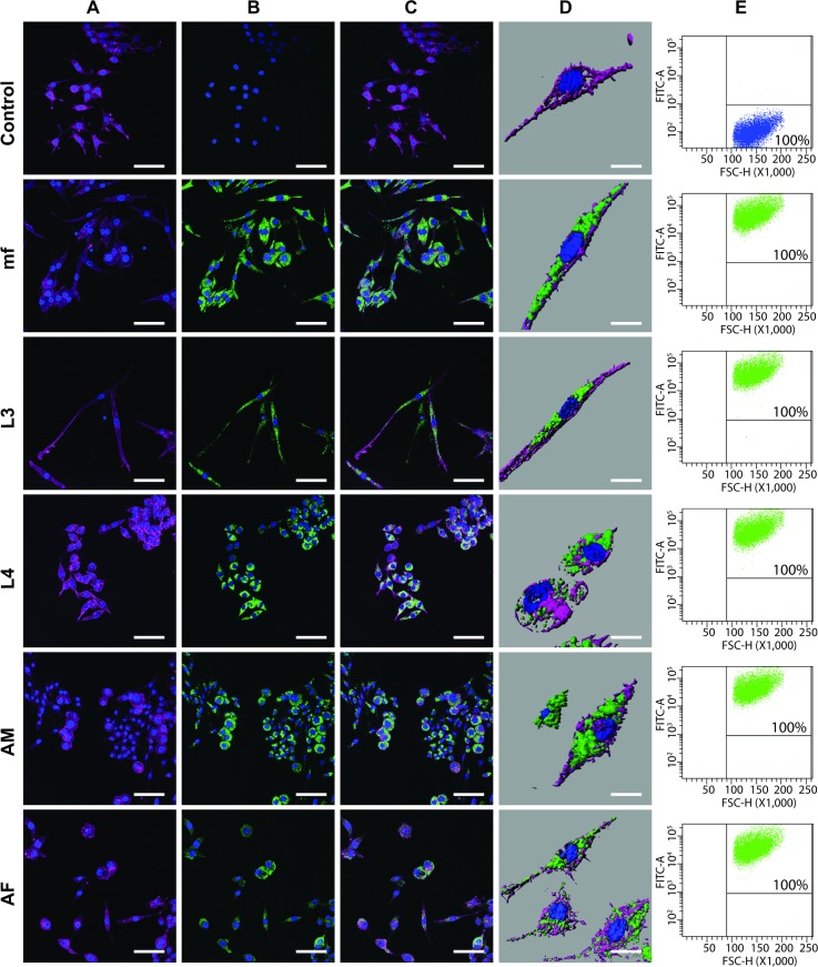
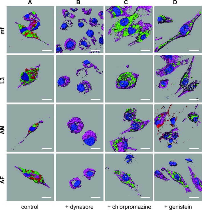
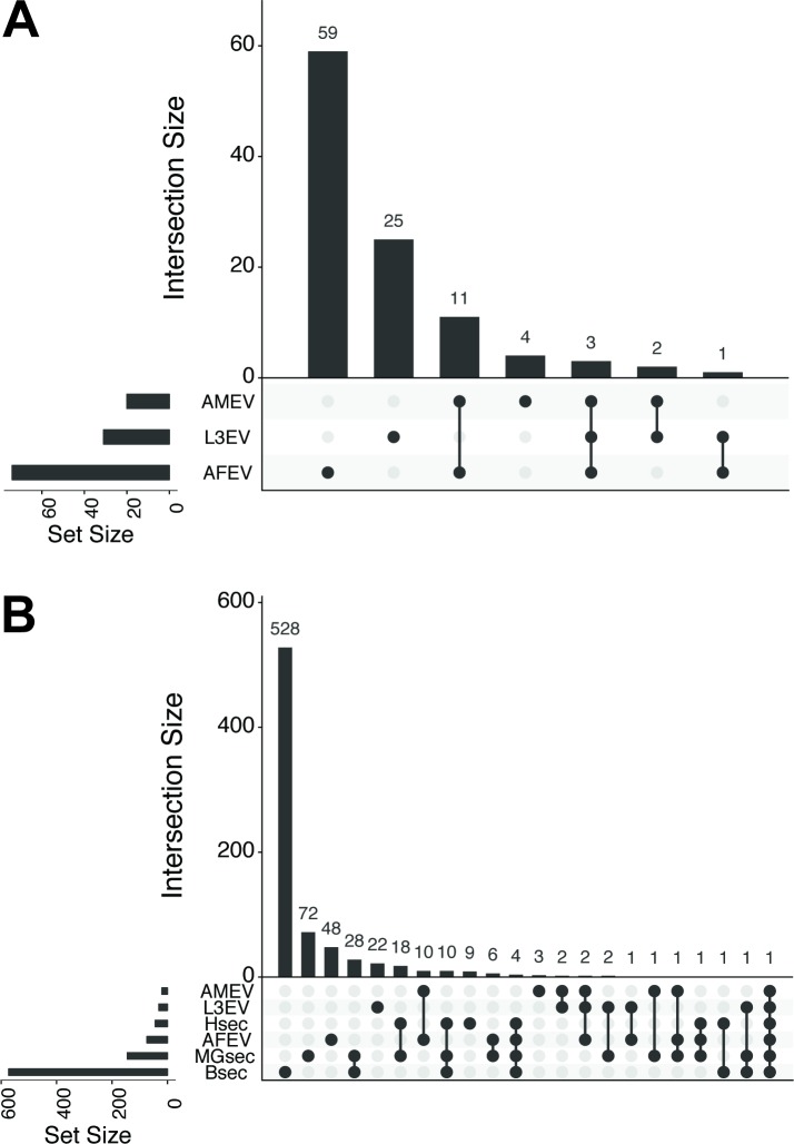
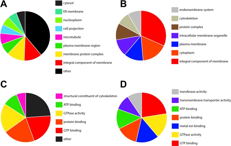
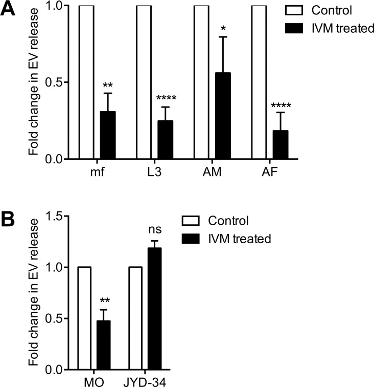
The filarial nematode Brugia malayi is an etiological agent of Lymphatic Filariasis. The capability of B. malayi and other parasitic nematodes to modulate host biology is recognized but the mechanisms by which such manipulation occurs are obscure. An emerging paradigm is the release of parasite-derived extracellular vesicles (EV) containing bioactive proteins and small RNA species that allow secretion of parasite effector molecules and their potential trafficking to host tissues. We have previously described EV release from the infectious L3 stage B. malayi and here we profile vesicle release across all intra-mammalian life cycle stages (microfilariae, L3, L4, adult male and female worms). Nanoparticle Tracking Analysis was used to quantify and size EVs revealing discrete vesicle populations and indicating a secretory process that is conserved across the life cycle. Brugia EVs are internalized by murine macrophages with no preference for life stage suggesting a uniform mechanism for effector molecule trafficking. Further, the use of chemical uptake inhibitors suggests all life stage EVs are internalized by phagocytosis. Proteomic profiling of adult male and female EVs using nano-scale LC-MS/MS described quantitative and qualitative differences in the adult EV proteome, helping define the biogenesis of Brugia EVs and revealing sexual dimorphic characteristics in immunomodulatory cargo. Finally, ivermectin was found to rapidly inhibit EV release by all Brugia life stages. Further this drug effect was also observed in the related filarial nematode, the canine heartworm Dirofilaria immitis but not in an ivermectin-unresponsive field isolate of that parasite, highlighting a potential mechanism of action for this drug and suggesting new screening platforms for anti-filarial drug development.
班氏吴策线虫是淋巴丝虫病的病原体。班氏吴策线虫和其他寄生线虫调节宿主生物学的能力已得到认可,但这种操纵发生的机制尚不清楚。一个新兴的范例是寄生虫衍生的细胞外囊泡(EV)的释放,其中包含生物活性蛋白和小 RNA 种类,允许寄生虫效应分子的分泌及其向宿主组织的潜在运输。我们之前已经描述了传染性 L3 期班氏吴策线虫的 EV 释放,在这里我们对所有哺乳动物生命周期阶段(微丝蚴、L3、L4、雌雄成虫)的囊泡释放进行了分析。纳米颗粒跟踪分析用于定量和大小 EV,揭示离散的囊泡群体,并表明分泌过程在整个生命周期中是保守的。Brugia EV 被鼠巨噬细胞内化,与生命阶段无关,这表明效应分子运输的机制是一致的。此外,使用化学摄取抑制剂表明,所有生命阶段的 EV 都是通过吞噬作用内化的。使用纳米级 LC-MS/MS 对成年雄性和雌性 EV 的蛋白质组学进行分析,描述了成年 EV 蛋白质组的定量和定性差异,有助于定义 Brugia EV 的生物发生,并揭示免疫调节货物的性别二态特征。最后,发现伊维菌素可迅速抑制所有班氏线虫生命周期阶段的 EV 释放。此外,这种药物效应也在相关的丝虫科线虫犬心丝虫中观察到,但在该寄生虫的一个对伊维菌素无反应的现场分离株中未观察到,这突出了该药物的潜在作用机制,并为抗丝虫药物开发提供了新的筛选平台。