Departamento de Bioquímica e Imunologia, Instituto de Ciências Biológicas, Universidade Federal de Minas Gerais, Belo Horizonte, Minas Gerais, Brazil.
Centro de Pesquisa e Desenvolvimento, Fundação Ezequiel Dias, Belo Horizonte, Minas Gerais, Brazil.
PLoS Negl Trop Dis. 2018 Apr 16;12(4):e0006427. doi: 10.1371/journal.pntd.0006427. eCollection 2018 Apr.
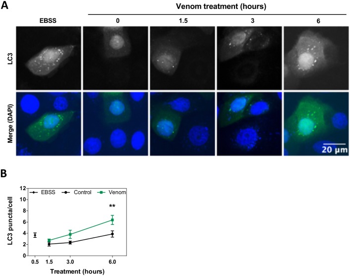
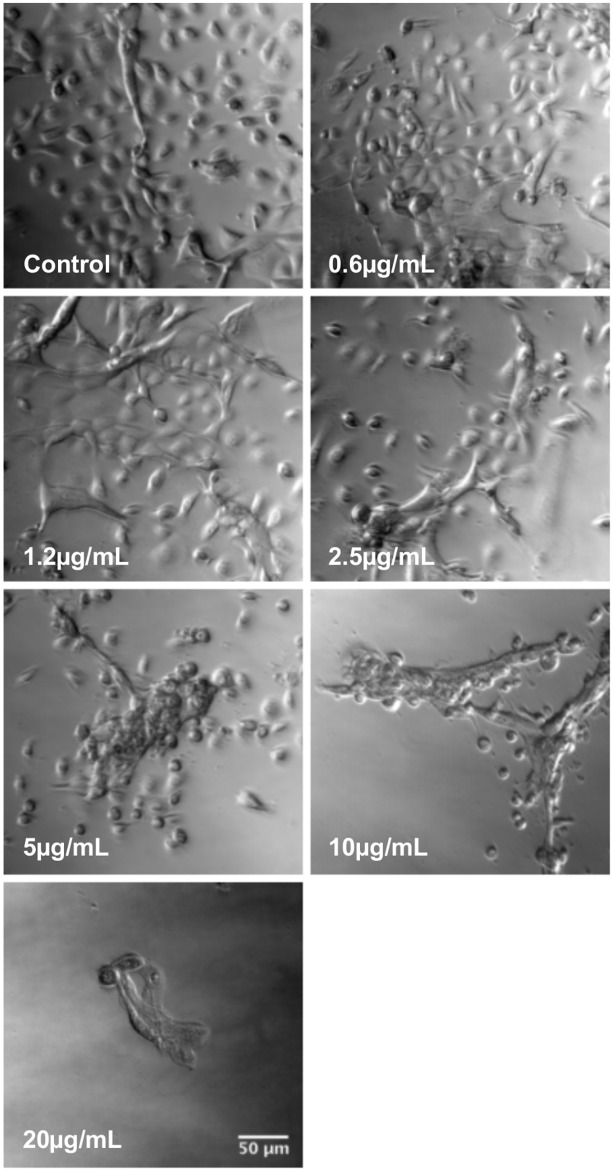
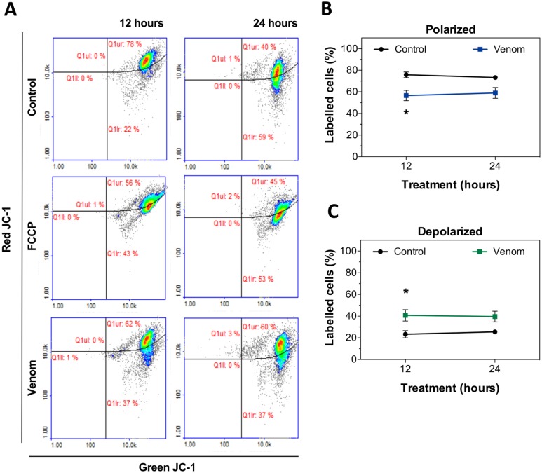
Envenomation by the bushmaster snake Lachesis muta muta is considered severe, characterized by local effects including necrosis, the main cause of permanent disability. However, cellular mechanisms related to cell death and tissue destruction, triggered by snake venoms, are poorly explored. The purpose of this study was to investigate the cytotoxic effect caused by L. m. muta venom in normal human keratinocytes and to identify the cellular processes involved in in cellulo envenomation. In order to investigate venom effect on different cell types, Alamar Blue assay was performed to quantify levels of cellular metabolism as a readout of cell viability. Apoptosis, necrosis and changes in mitochondrial membrane potential were evaluated by flow cytometry, while induction of autophagy was assessed by expression of GFP-LC3 and analyzed using fluorescence microscopy. The cytotoxic potential of the venom is shown by reduced cell viability in a concentration-dependent manner. It was also observed the sequential appearance of cells undergoing autophagy (by 6 hours), apoptosis and necrosis (12 and 24 hours). Morphologically, incubation with L. m. muta venom led to a significant cellular retraction and formation of cellular aggregates. These results indicate that L. m. muta venom is cytotoxic to normal human keratinocytes and other cell lines, and this toxicity involves the integration of distinct modes of cell death. Autophagy as a cell death mechanism, in addition to apoptosis and necrosis, can help to unravel cellular pathways and mechanisms triggered by the venom. Understanding the mechanisms that underlie cellular damage and tissue destruction will be useful in the development of alternative therapies against snakebites.
被矛头蝮蛇(Lachesis muta muta)咬伤被认为是严重的,其特征是局部效应,包括坏死,这是永久性残疾的主要原因。然而,蛇毒引发的细胞死亡和组织破坏的细胞机制还未得到充分探索。本研究旨在研究矛头蝮蛇毒液对正常人类角质形成细胞的细胞毒性作用,并确定与细胞内中毒相关的细胞过程。为了研究毒液对不同细胞类型的影响,通过阿尔玛蓝测定法(Alamar Blue assay)来定量细胞代谢水平,作为细胞活力的读数。通过流式细胞术评估细胞凋亡、坏死和线粒体膜电位的变化,而自噬的诱导则通过 GFP-LC3 的表达进行评估,并使用荧光显微镜进行分析。毒液的细胞毒性潜力表现为细胞活力在浓度依赖性方式下降低。还观察到细胞依次经历自噬(在 6 小时)、凋亡和坏死(在 12 小时和 24 小时)。孵育矛头蝮蛇毒液后,细胞形态发生明显的回缩,并形成细胞聚集。这些结果表明,矛头蝮蛇毒液对正常人类角质形成细胞和其他细胞系具有细胞毒性,这种毒性涉及不同细胞死亡方式的整合。自噬作为一种细胞死亡机制,除了凋亡和坏死外,还可以帮助阐明毒液引发的细胞通路和机制。了解导致细胞损伤和组织破坏的机制将有助于开发针对蛇咬伤的替代疗法。