Morrison Robert J, Nasser Hassan B, Kashlan Khaled N, Zopf David A, Milner Derek J, Flanangan Colleen L, Wheeler Matthew B, Green Glenn E, Hollister Scott J
Department of Otolaryngology-Head and Neck Surgery, Vanderbilt University, Nashville, Tennessee.
Department of Otolaryngology-Head and Neck Surgery, University of California Los Angeles, Los Angeles, California.
Laryngoscope. 2018 Jul;128(7):E251-E257. doi: 10.1002/lary.27200. Epub 2018 Apr 18.
OBJECTIVES/HYPOTHESIS: Reconstruction of craniofacial cartilagenous defects are among the most challenging surgical procedures in facial plastic surgery. Bioengineered craniofacial cartilage holds immense potential to surpass current reconstructive options, but limitations to clinical translation exist. We endeavored to determine the viability of utilizing adipose-derived stem cell-chondrocyte co-culture and three-dimensional (3D) printing to produce 3D bioscaffolds for cartilage tissue engineering. We describe a feasibility study revealing a novel approach for cartilage tissue engineering with in vitro and in vivo animal data.
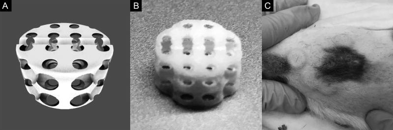
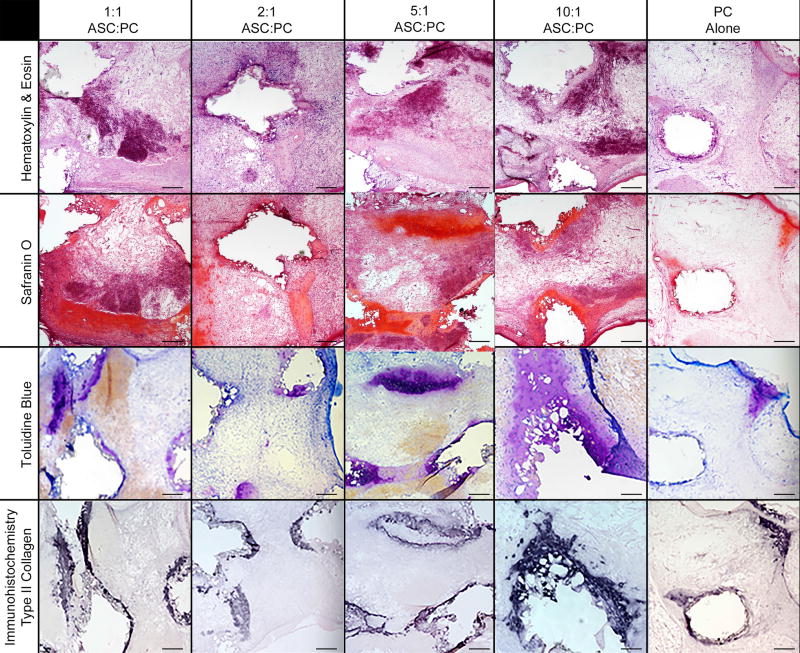
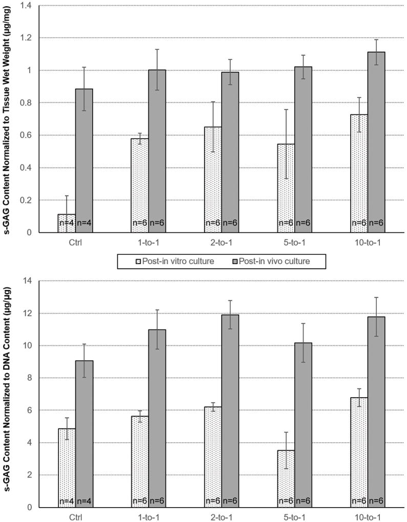
Porcine adipose-derived stem cells and chondrocytes were isolated and co-seeded at 1:1, 2:1, 5:1, 10:1, and 0:1 experimental ratios in a hyaluronic acid/collagen hydrogel in the pores of 3D-printed polycaprolactone scaffolds to form 3D bioscaffolds for cartilage tissue engineering. Bioscaffolds were cultured in vitro without growth factors for 4 weeks and then implanted into the subcutaneous tissue of athymic rats for an additional 4 weeks before sacrifice. Bioscaffolds were subjected to histologic, immunohistochemical, and biochemical analysis.
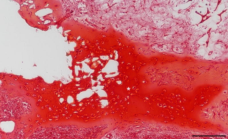
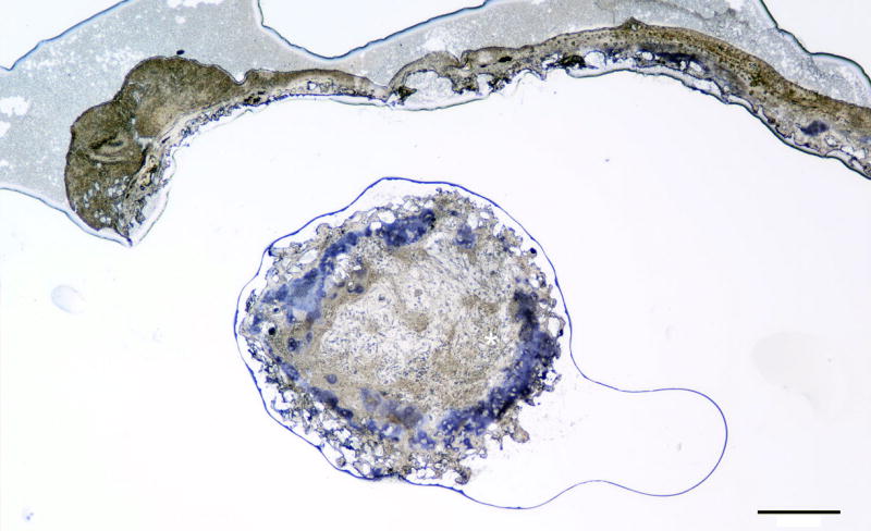
Successful production of cartilage was achieved using a co-culture model of adipose-derived stem cells and chondrocytes without the use of exogenous growth factors. Histology demonstrated cartilage growth for all experimental ratios at the post-in vivo time point confirmed with type II collagen immunohistochemistry. There was no difference in sulfated-glycosaminoglycan production between experimental groups.
Tissue-engineered cartilage was successfully produced on 3D-printed bioresorbable scaffolds using an adipose-derived stem cell and chondrocyte co-culture technique. This potentiates co-culture as a solution for several key barriers to a clinically translatable cartilage tissue engineering process.
NA. Laryngoscope, 128:E251-E257, 2018.
目的/假设:颅面软骨缺损的重建是面部整形手术中最具挑战性的外科手术之一。生物工程化的颅面软骨具有巨大潜力,有望超越现有的重建方法,但在临床转化方面存在局限性。我们致力于确定利用脂肪干细胞 - 软骨细胞共培养和三维(3D)打印来生产用于软骨组织工程的3D生物支架的可行性。我们描述了一项可行性研究,该研究通过体外和体内动物数据揭示了一种软骨组织工程的新方法。
分离猪脂肪干细胞和软骨细胞,并以1:1、2:1、5:1、10:1和0:1的实验比例共接种于3D打印的聚己内酯支架孔隙中的透明质酸/胶原蛋白水凝胶中,以形成用于软骨组织工程的3D生物支架。生物支架在无生长因子的条件下体外培养4周,然后植入无胸腺大鼠的皮下组织中再培养4周后处死。对生物支架进行组织学、免疫组织化学和生化分析。
使用脂肪干细胞和软骨细胞的共培养模型,无需使用外源性生长因子即可成功生成软骨。组织学显示,在体内后时间点,所有实验比例均有软骨生长,经II型胶原免疫组织化学证实。各实验组之间硫酸化糖胺聚糖的产生没有差异。
使用脂肪干细胞和软骨细胞共培养技术,在3D打印的生物可吸收支架上成功制备了组织工程软骨。这增强了共培养作为解决临床可转化软骨组织工程过程中几个关键障碍的一种方法的潜力。
无。《喉镜》,2018年,第128卷,E251 - E257页。