• 文献检索
  • 文档翻译
  • 深度研究
  • 学术资讯
  • Suppr Zotero 插件Zotero 插件
  • 邀请有礼
  • 套餐&价格
  • 历史记录
应用&插件
Suppr Zotero 插件Zotero 插件浏览器插件Mac 客户端Windows 客户端微信小程序
定价
高级版会员购买积分包购买API积分包
服务
文献检索文档翻译深度研究API 文档MCP 服务
关于我们
关于 Suppr公司介绍联系我们用户协议隐私条款
关注我们

Suppr 超能文献

核心技术专利:CN118964589B侵权必究
粤ICP备2023148730 号-1Suppr @ 2026

文献检索

告别复杂PubMed语法,用中文像聊天一样搜索,搜遍4000万医学文献。AI智能推荐,让科研检索更轻松。

立即免费搜索

文件翻译

保留排版,准确专业,支持PDF/Word/PPT等文件格式,支持 12+语言互译。

免费翻译文档

深度研究

AI帮你快速写综述,25分钟生成高质量综述,智能提取关键信息,辅助科研写作。

立即免费体验

利用细胞器靶向分割 GFP 系统可视化多个细胞器间接触位点。

Visualizing multiple inter-organelle contact sites using the organelle-targeted split-GFP system.

机构信息

Faculty of Science, Yamagata University, 1-4-12 Kojirakawa-machi, Yamagata, Yamagata, 990-8560, Japan.

Faculty of Life Sciences, Kyoto Sangyo University, Kamigamo-motoyama, Kita-ku, Kyoto, 603-8555, Japan.

出版信息

Sci Rep. 2018 Apr 18;8(1):6175. doi: 10.1038/s41598-018-24466-0.

DOI:10.1038/s41598-018-24466-0
PMID:29670150
原文链接:https://pmc.ncbi.nlm.nih.gov/articles/PMC5906596/
Abstract

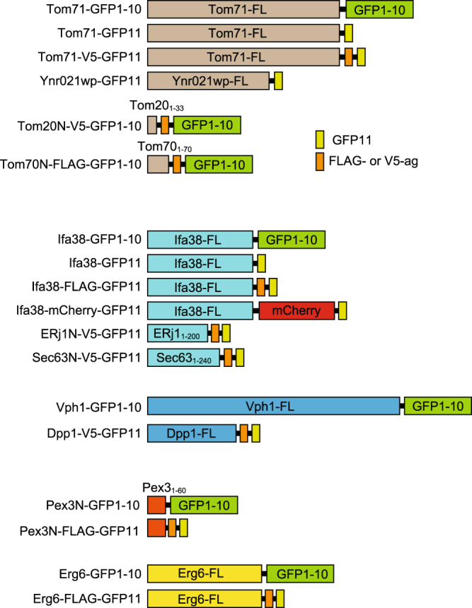
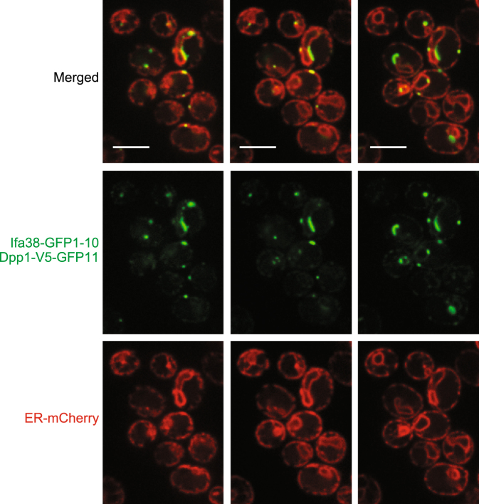
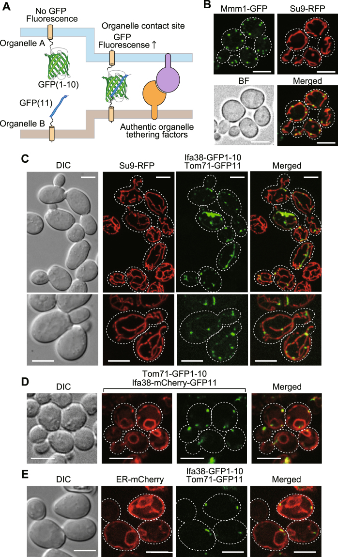
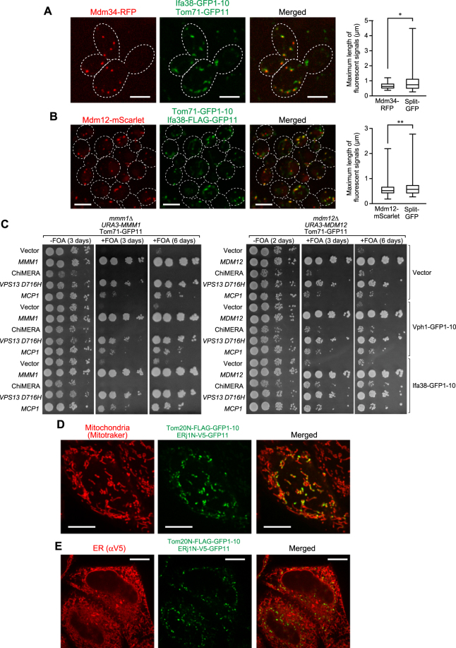
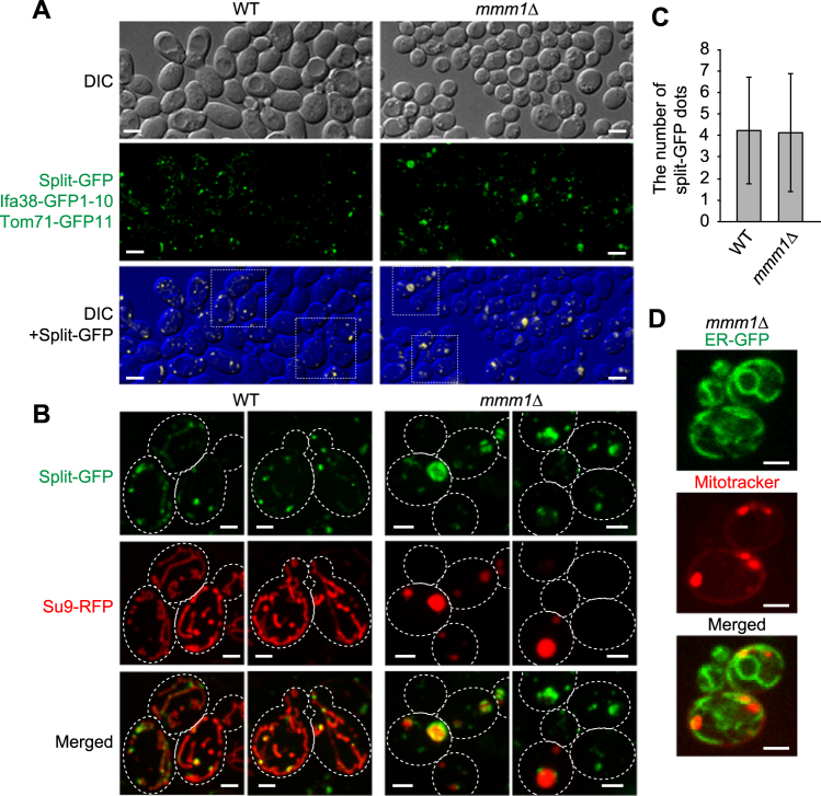
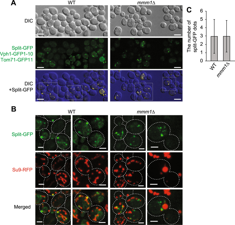
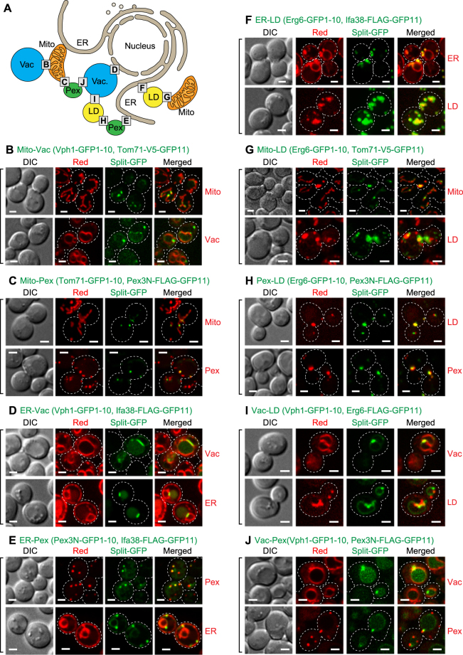
Functional integrity of eukaryotic organelles relies on direct physical contacts between distinct organelles. However, the entity of organelle-tethering factors is not well understood due to lack of means to analyze inter-organelle interactions in living cells. Here we evaluate the split-GFP system for visualizing organelle contact sites in vivo and show its advantages and disadvantages. We observed punctate GFP signals from the split-GFP fragments targeted to any pairs of organelles among the ER, mitochondria, peroxisomes, vacuole and lipid droplets in yeast cells, which suggests that these organelles form contact sites with multiple organelles simultaneously although it is difficult to rule out the possibilities that these organelle contacts sites are artificially formed by the irreversible associations of the split-GFP probes. Importantly, split-GFP signals in the overlapped regions of the ER and mitochondria were mainly co-localized with ERMES, an authentic ER-mitochondria tethering structure, suggesting that split-GFP assembly depends on the preexisting inter-organelle contact sites. We also confirmed that the split-GFP system can be applied to detection of the ER-mitochondria contact sites in HeLa cells. We thus propose that the split-GFP system is a potential tool to observe and analyze inter-organelle contact sites in living yeast and mammalian cells.

摘要

真核细胞器的功能完整性依赖于不同细胞器之间的直接物理接触。然而,由于缺乏在活细胞中分析细胞器相互作用的手段,因此对细胞器连接因子的实体并不了解。在这里,我们评估了用于在体内可视化细胞器接触位点的分裂 GFP 系统,并展示了其优缺点。我们观察到来自酵母细胞中内质网、线粒体、过氧化物酶体、液泡和脂滴之间任何两对细胞器的靶向的分裂 GFP 片段的点状 GFP 信号,这表明尽管很难排除这些细胞器接触位点是由分裂 GFP 探针的不可逆结合人工形成的可能性,但这些细胞器同时与多个细胞器形成接触位点。重要的是,内质网和线粒体的重叠区域中的分裂 GFP 信号主要与 ERMES 共定位,后者是一种真实的内质网-线粒体连接结构,这表明分裂 GFP 的组装取决于预先存在的细胞器之间的接触位点。我们还证实,该分裂 GFP 系统可用于检测 HeLa 细胞中的 ER-线粒体接触位点。因此,我们提出分裂 GFP 系统是一种在活酵母和哺乳动物细胞中观察和分析细胞器接触位点的潜在工具。

https://cdn.ncbi.nlm.nih.gov/pmc/blobs/9e92/5906596/b92b8e9d5540/41598_2018_24466_Fig7_HTML.jpg
https://cdn.ncbi.nlm.nih.gov/pmc/blobs/9e92/5906596/7a5bfeb10618/41598_2018_24466_Fig1_HTML.jpg
https://cdn.ncbi.nlm.nih.gov/pmc/blobs/9e92/5906596/041e25d1dbd4/41598_2018_24466_Fig2_HTML.jpg
https://cdn.ncbi.nlm.nih.gov/pmc/blobs/9e92/5906596/fa250a875b47/41598_2018_24466_Fig3_HTML.jpg
https://cdn.ncbi.nlm.nih.gov/pmc/blobs/9e92/5906596/c28cd6c9b638/41598_2018_24466_Fig4_HTML.jpg
https://cdn.ncbi.nlm.nih.gov/pmc/blobs/9e92/5906596/55d7a854ce7d/41598_2018_24466_Fig5_HTML.jpg
https://cdn.ncbi.nlm.nih.gov/pmc/blobs/9e92/5906596/487de4174b22/41598_2018_24466_Fig6_HTML.jpg
https://cdn.ncbi.nlm.nih.gov/pmc/blobs/9e92/5906596/b92b8e9d5540/41598_2018_24466_Fig7_HTML.jpg

相似文献

1
Visualizing multiple inter-organelle contact sites using the organelle-targeted split-GFP system.利用细胞器靶向分割 GFP 系统可视化多个细胞器间接触位点。
Sci Rep. 2018 Apr 18;8(1):6175. doi: 10.1038/s41598-018-24466-0.
2
Improved Split-GFP Systems for Visualizing Organelle Contact Sites in Yeast and Human Cells.用于可视化酵母和人类细胞中细胞器接触位点的改良型分裂绿色荧光蛋白系统
Front Cell Dev Biol. 2020 Nov 20;8:571388. doi: 10.3389/fcell.2020.571388. eCollection 2020.
3
Cellular metabolism regulates contact sites between vacuoles and mitochondria.细胞代谢调节液泡和线粒体之间的接触部位。
Dev Cell. 2014 Jul 14;30(1):86-94. doi: 10.1016/j.devcel.2014.06.006.
4
Organelle contact zones as sites for lipid transfer.细胞器接触区作为脂质转移的位点。
J Biochem. 2019 Feb 1;165(2):115-123. doi: 10.1093/jb/mvy088.
5
Split Green Fluorescent Protein-Based Contact Site Sensor (SPLICS ) for Heterotypic Organelle Juxtaposition as Applied to ER -Mitochondria Proximities.基于分裂绿色荧光蛋白的接触位点传感器(SPLICS)用于异型细胞器并置,应用于 ER-线粒体接近。
Methods Mol Biol. 2021;2275:363-378. doi: 10.1007/978-1-0716-1262-0_23.
6
ER membranes exhibit phase behavior at sites of organelle contact.内质网膜在细胞器接触部位表现出相行为。
Proc Natl Acad Sci U S A. 2020 Mar 31;117(13):7225-7235. doi: 10.1073/pnas.1910854117. Epub 2020 Mar 16.
7
Role of Intra- and Inter-mitochondrial Membrane Contact Sites in Yeast Phospholipid Biogenesis.酵母磷脂生物合成中线粒体内外膜接触位点的作用
Adv Exp Med Biol. 2017;997:121-133. doi: 10.1007/978-981-10-4567-7_9.
8
Inter-organelle membrane contact sites: implications for lipid metabolism.细胞器间膜接触位点:对脂质代谢的影响。
Biol Direct. 2020 Nov 11;15(1):24. doi: 10.1186/s13062-020-00279-y.
9
A Combinatorial Reporter Set to Visualize the Membrane Contact Sites Between Endoplasmic Reticulum and Other Organelles in Plant Cell.用于可视化植物细胞内质网与其他细胞器之间膜接触位点的组合报告基因集
Front Plant Sci. 2020 Aug 18;11:1280. doi: 10.3389/fpls.2020.01280. eCollection 2020.
10
Applying systems-level spectral imaging and analysis to reveal the organelle interactome.应用系统级光谱成像和分析来揭示细胞器相互作用组。
Nature. 2017 Jun 1;546(7656):162-167. doi: 10.1038/nature22369. Epub 2017 May 24.

引用本文的文献

1
Dnm1 Is Required for the Focal Clustering of Fis1 on the Mitochondrial Outer Membrane.Dnm1是线粒体外膜上Fis1焦点聚集所必需的。
MicroPubl Biol. 2025 Aug 19;2025. doi: 10.17912/micropub.biology.001780. eCollection 2025.
2
Pex3 promotes formation of peroxisome-peroxisome and peroxisome-lipid droplet contact sites.Pex3促进过氧化物酶体-过氧化物酶体以及过氧化物酶体-脂滴接触位点的形成。
Sci Rep. 2025 Jul 8;15(1):24480. doi: 10.1038/s41598-025-07934-2.
3
Efficient endogenous protein labelling in Dictyostelium using CRISPR/Cas9 knock-in and split fluorescent proteins.

本文引用的文献

1
Vps13-Mcp1 interact at vacuole-mitochondria interfaces and bypass ER-mitochondria contact sites.Vps13与Mcp1在液泡-线粒体界面相互作用,并绕过内质网-线粒体接触位点。
J Cell Biol. 2017 Oct 2;216(10):3219-3229. doi: 10.1083/jcb.201610055. Epub 2017 Sep 1.
2
Function of lipid droplet-organelle interactions in lipid homeostasis.脂滴-细胞器相互作用在脂质动态平衡中的功能。
Biochim Biophys Acta Mol Cell Res. 2017 Sep;1864(9):1459-1468. doi: 10.1016/j.bbamcr.2017.04.001. Epub 2017 Apr 5.
3
A Tether Is a Tether Is a Tether: Tethering at Membrane Contact Sites.
利用CRISPR/Cas9敲入和分裂荧光蛋白在盘基网柄菌中进行高效内源性蛋白质标记
PLoS One. 2025 Jun 20;20(6):e0326577. doi: 10.1371/journal.pone.0326577. eCollection 2025.
4
Split genetically encoded calcium indicators for interorganellar junctions.用于细胞器间连接的分裂型基因编码钙指示剂。
Proc Natl Acad Sci U S A. 2025 May 20;122(20):e2415268122. doi: 10.1073/pnas.2415268122. Epub 2025 May 13.
5
Various methods to detect small GTPase activation: from radioisotope-based methods to the Small GTPase ActIvitY ANalysing (SAIYAN) system.检测小GTP酶激活的各种方法:从基于放射性同位素的方法到小GTP酶活性分析(SAIYAN)系统。
J Biochem. 2025 Apr 29;177(5):321-327. doi: 10.1093/jb/mvaf012.
6
Split-Luciferase Reassembly Assay to Measure Endoplasmic Reticulum-Mitochondria Contacts in Live Cells.分裂萤光素酶重组分析测定活细胞内质网-线粒体接触。
J Vis Exp. 2024 Oct 11(212). doi: 10.3791/66862.
7
Imaging and proteomics toolkits for studying organelle contact sites.用于研究细胞器接触位点的成像和蛋白质组学工具包。
Front Cell Dev Biol. 2024 Sep 24;12:1466915. doi: 10.3389/fcell.2024.1466915. eCollection 2024.
8
Small GTPase ActIvitY ANalyzing (SAIYAN) system: A method to detect GTPase activation in living cells.小 GTPase 活性分析(SAIYAN)系统:一种检测活细胞中 GTPase 激活的方法。
J Cell Biol. 2024 Oct 7;223(10). doi: 10.1083/jcb.202403179. Epub 2024 Aug 5.
9
A fluorogenic complementation tool kit for interrogating lipid droplet-organelle interaction.用于研究脂滴-细胞器相互作用的荧光互补工具包。
J Cell Biol. 2024 Sep 2;223(9). doi: 10.1083/jcb.202311126. Epub 2024 Jul 1.
10
Mitochondria-ER-PM contacts regulate mitochondrial division and PI(4)P distribution.线粒体-内质网-质膜接触调控线粒体分裂和 PI(4)P 分布。
J Cell Biol. 2024 Sep 2;223(9). doi: 10.1083/jcb.202308144. Epub 2024 May 23.
A Tether Is a Tether Is a Tether: Tethering at Membrane Contact Sites. 系绳就是系绳就是系绳:在膜接触位点系绳。
Dev Cell. 2016 Nov 21;39(4):395-409. doi: 10.1016/j.devcel.2016.10.022.
4
mScarlet: a bright monomeric red fluorescent protein for cellular imaging.mScarlet:一种明亮的单体红色荧光蛋白,用于细胞成像。
Nat Methods. 2017 Jan;14(1):53-56. doi: 10.1038/nmeth.4074. Epub 2016 Nov 21.
5
Identification of multi-copy suppressors for endoplasmic reticulum-mitochondria tethering proteins in Saccharomyces cerevisiae.酿酒酵母中内质网-线粒体锚定蛋白多拷贝抑制子的鉴定。
FEBS Lett. 2016 Sep;590(18):3061-70. doi: 10.1002/1873-3468.12358. Epub 2016 Sep 1.
6
A phospholipid transfer function of ER-mitochondria encounter structure revealed in vitro.体外揭示的内质网-线粒体接触结构的磷脂转移功能。
Sci Rep. 2016 Jul 29;6:30777. doi: 10.1038/srep30777.
7
Vacuole membrane contact sites and domains: emerging hubs to coordinate organelle function with cellular metabolism.液泡膜接触位点与区域:协调细胞器功能与细胞代谢的新兴枢纽
Biochem Soc Trans. 2016 Apr 15;44(2):528-33. doi: 10.1042/BST20150277.
8
Lipid transfer proteins do their thing anchored at membrane contact sites… but what is their thing?脂质转运蛋白在膜接触位点发挥作用……但它们的作用是什么呢?
Biochem Soc Trans. 2016 Apr 15;44(2):517-27. doi: 10.1042/BST20150275.
9
ER-mitochondrial junctions can be bypassed by dominant mutations in the endosomal protein Vps13.内体蛋白Vps13中的显性突变可绕过内质网-线粒体连接点。
J Cell Biol. 2015 Sep 14;210(6):883-90. doi: 10.1083/jcb.201502105.
10
Mdm1/Snx13 is a novel ER-endolysosomal interorganelle tethering protein.Mdm1/Snx13是一种新型的内质网-内溶酶体细胞器间连接蛋白。
J Cell Biol. 2015 Aug 17;210(4):541-51. doi: 10.1083/jcb.201503088.