Vascular Biology Center, Medical College of Georgia, Augusta University, Augusta, GA 30912, USA.
Charlie Norwood VA Medical Center, Augusta, GA 30904, USA.
Int J Mol Sci. 2018 Apr 17;19(4):1215. doi: 10.3390/ijms19041215.
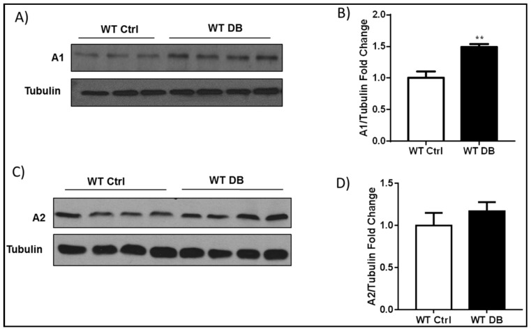
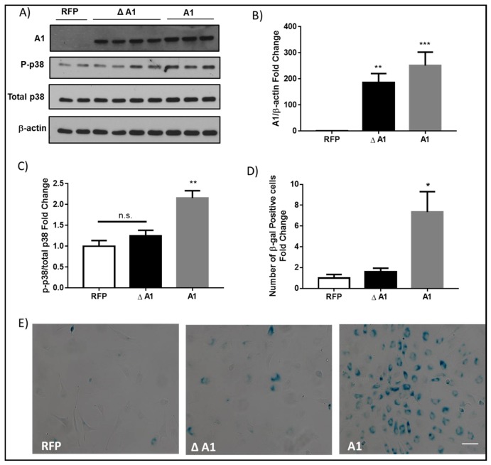
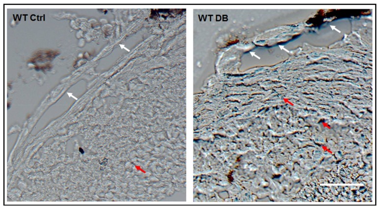
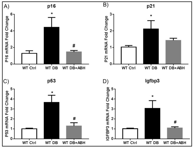
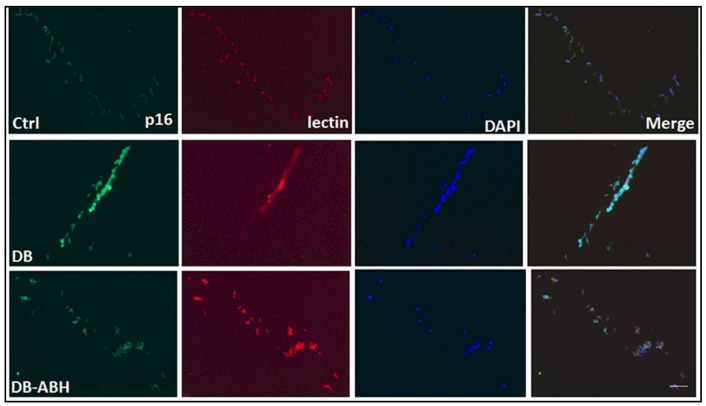
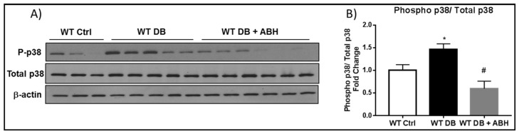
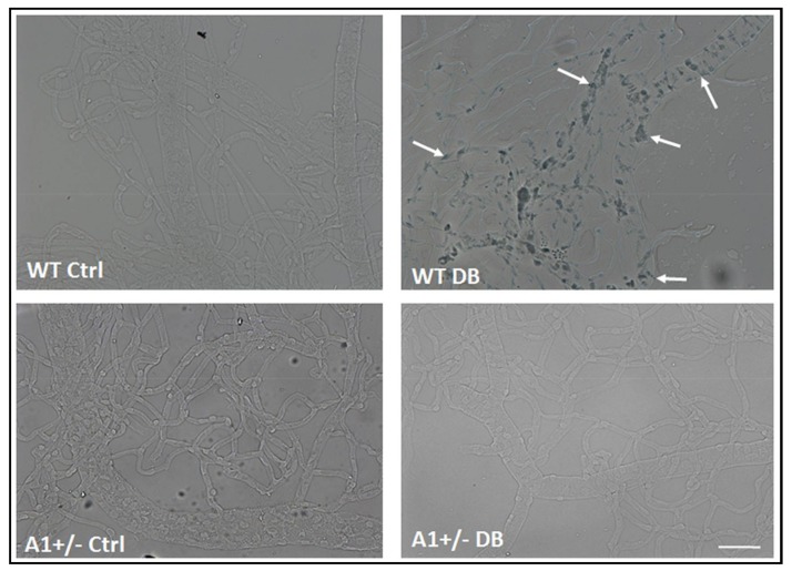
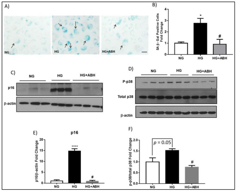
We have recently found that diabetes-induced premature senescence of retinal endothelial cells is accompanied by NOX2-NADPH oxidase-induced increases in the ureohydrolase enzyme arginase 1 (A1). Here, we used genetic strategies to determine the specific involvement of A1 in diabetes-induced endothelial cell senescence. We used A1 knockout mice and wild type mice that were rendered diabetic with streptozotocin and retinal endothelial cells (ECs) exposed to high glucose or transduced with adenovirus to overexpress A1 for these experiments. ABH [2(S)-Amino-6-boronohexanoic acid] was used to inhibit arginase activity. We used Western blotting, immunolabeling, quantitative PCR, and senescence associated β-galactosidase (SA β-Gal) activity to evaluate senescence. Analyses of retinal tissue extracts from diabetic mice showed significant increases in mRNA expression of the senescence-related proteins p16, p21, and p53 when compared with non-diabetic mice. SA β-Gal activity and p16 immunoreactivity were also increased in retinal vessels from diabetic mice. A1 gene deletion or pharmacological inhibition protected against the induction of premature senescence. A1 overexpression or high glucose treatment increased SA β-Gal activity in cultured ECs. These results demonstrate that A1 is critically involved in diabetes-induced senescence of retinal ECs. Inhibition of arginase activity may therefore be an effective therapeutic strategy to alleviate diabetic retinopathy by preventing premature senescence.
我们最近发现,糖尿病引起的视网膜内皮细胞过早衰老伴随着 NADPH 氧化酶诱导的尿水解酶精氨酸酶 1(A1)增加。在这里,我们使用遗传策略来确定 A1 在糖尿病诱导的内皮细胞衰老中的具体作用。我们使用 A1 敲除小鼠和野生型小鼠,用链脲佐菌素使这些小鼠患上糖尿病,并使视网膜内皮细胞(EC)暴露于高葡萄糖或用腺病毒转导以过表达 A1 进行这些实验。ABH [2(S)-氨基-6-硼己酸] 用于抑制精氨酸酶活性。我们使用 Western blot、免疫标记、定量 PCR 和衰老相关的 β-半乳糖苷酶(SA β-Gal)活性来评估衰老。与非糖尿病小鼠相比,糖尿病小鼠的视网膜组织提取物分析显示衰老相关蛋白 p16、p21 和 p53 的 mRNA 表达显著增加。视网膜血管中的 SA β-Gal 活性和 p16 免疫反应性也增加。A1 基因缺失或药理学抑制可防止过早衰老的诱导。A1 过表达或高葡萄糖处理增加了培养的 EC 中的 SA β-Gal 活性。这些结果表明,A1 在内皮细胞糖尿病诱导的衰老中起着关键作用。因此,抑制精氨酸酶活性可能是通过防止过早衰老来减轻糖尿病性视网膜病变的有效治疗策略。