Athinoula A. Martinos Center for Biomedical Imaging, Massachusetts General Hospital, Charlestown, Massachusetts.
Fetal-Neonatal Neuroimaging and Developmental Science Center, Division of Newborn Medicine, Department of Medicine, Boston Children's Hospital, Harvard Medical School, Boston, Massachusetts.
Pediatr Neurol. 2018 Jun;83:25-31. doi: 10.1016/j.pediatrneurol.2018.03.004. Epub 2018 Mar 15.
The purpose of this study is to clarify the source distribution patterns of magnetoencephalographic spikes correlated with postsurgical seizure-free outcome in pediatric patients with focal cortical dysplasia.
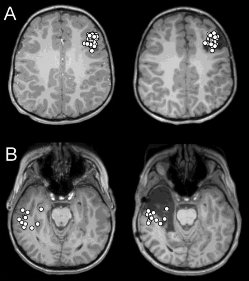
Thirty-two patients with pathologically confirmed focal cortical dysplasia were divided into seizure-free and seizure-persistent groups according to their surgical outcomes based on Engel classification. In each patient, presurgical magnetoencephalography was reviewed. Dipole sources of magnetoencephalographic spikes were calculated according to a single dipole model. We obtained the following quantitative indices for evaluating dipole distribution: maximum distance over all pairs of dipoles, standard deviation of the distances between each dipole and the mean coordinate of all dipoles, average nearest neighbor distance, the rate of dipoles located within 10, 20, and 30 mm from the mean coordinate, and the rate of dipoles included in the resection. These indices were compared between the two patient groups.
Average nearest neighbor distance was significantly smaller in the seizure-free group than in the seizure-persistent group (P = 0.008). The rates of dipoles located within 10, 20, and 30 mm from the mean coordinate were significantly higher in the seizure-free group (P = 0.001, 0.001, 0.005, respectively). The maximum distance, standard deviation, and resection rate of dipoles did not show a significant difference between the two groups.
A spatially restricted dipole distribution of magnetoencephalographic spikes is correlated with postsurgical seizure-free outcomes in patients with focal cortical dysplasia. The distribution can be assessed by quantitative indices that are clinically useful in the presurgical evaluation of these patients.
本研究旨在阐明与术后无癫痫发作结果相关的皮质发育不良患儿的磁源性影像棘波的源分布模式。
根据 Engel 分级,根据手术结果将 32 例经病理证实的局灶性皮质发育不良患者分为无癫痫发作组和癫痫持续组。对每位患者的术前磁源性影像进行回顾。根据单偶极子模型计算磁源性影像棘波的偶极子源。我们获得了以下用于评估偶极子分布的定量指标:所有偶极子对之间的最大距离、每个偶极子与所有偶极子的平均坐标之间的距离的标准差、平均最近邻距离、位于距平均坐标 10、20 和 30mm 以内的偶极子的比率以及位于切除范围内的偶极子的比率。将这些指标在两组患者之间进行比较。
无癫痫发作组的平均最近邻距离明显小于癫痫持续组(P=0.008)。位于距平均坐标 10、20 和 30mm 以内的偶极子的比率在无癫痫发作组中显著更高(P=0.001、0.001 和 0.005)。偶极子的最大距离、标准差和切除率在两组之间没有显著差异。
皮质发育不良患者的磁源性影像棘波的偶极子分布具有空间限制,与术后无癫痫发作结果相关。这些分布可以通过定量指标来评估,这些指标在这些患者的术前评估中具有临床意义。