Annu Int Conf IEEE Eng Med Biol Soc. 2021 Nov;2021:2668-2671. doi: 10.1109/EMBC46164.2021.9630246.
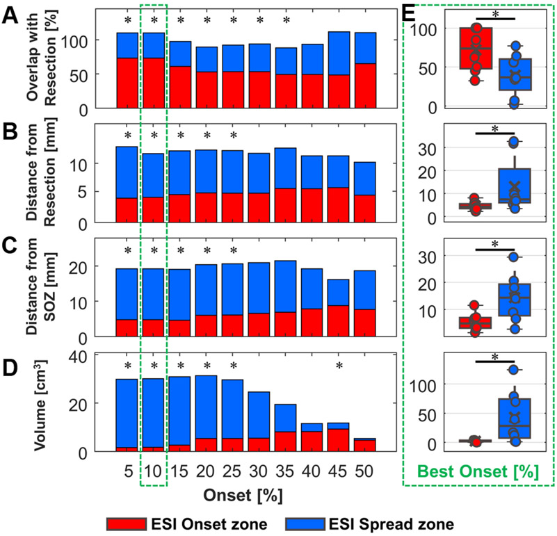
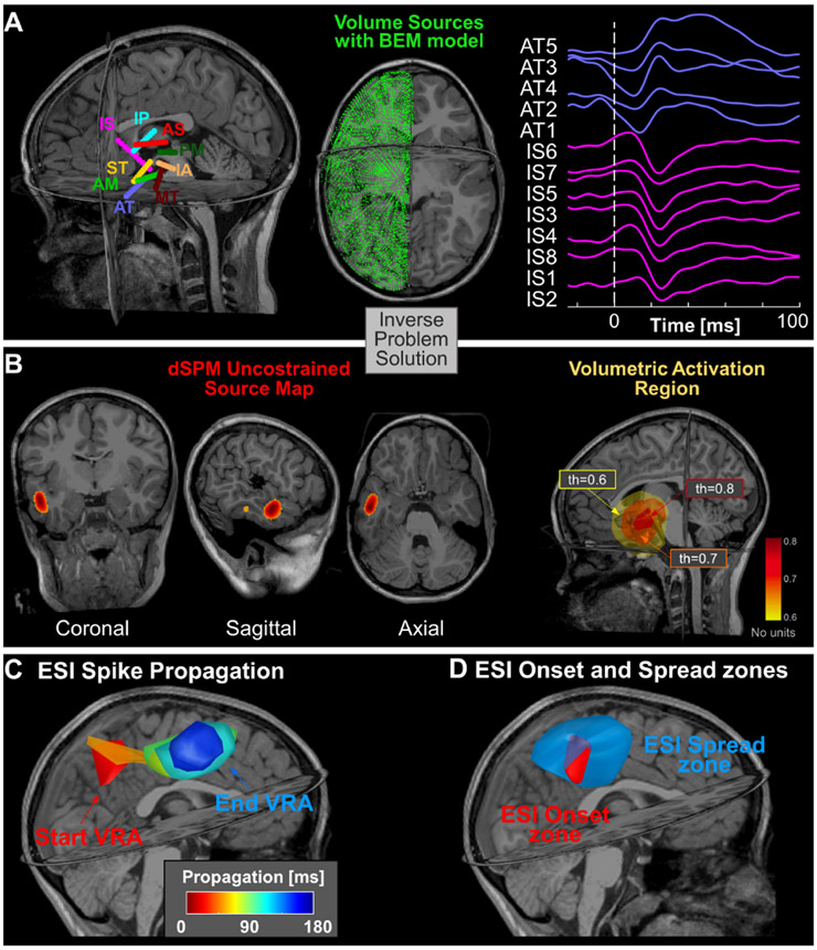
Interictal epileptiform discharges (IEDs) serve as sensitive but not specific biomarkers of epilepsy that can delineate the epileptogenic zone (EZ) in patients with drug resistant epilepsy (DRE) undergoing surgery. Intracranial EEG (icEEG) studies have shown that IEDs propagate in time across large areas of the brain. The onset of this propagation is regarded as a more specific biomarker of epilepsy than areas of spread. Yet, the limited spatial resolution of icEEG does not allow to identify the onset of this activity with high precision. Here, we propose a new method of mapping the spatiotemporal propagation of IEDs (and identify its onset) by using Electrical Source Imaging (ESI) on icEEG bypassing the spatial limitations of icEEG. We validated our method on icEEG recordings from 8 children with DRE who underwent surgery with good outcome (Engel score =1). On each icEEG channel, we detected IEDs and identified the propagation onset using an automated algorithm. We localized the propagation of IEDs with dynamic Statistical Parametric Mapping (dSPM) using a time-sliding window approach. We defined two brain regions: the ESI-onset and ESI-spread zone. We estimated the overlap of these regions with resection volume (in percentage), which served as the gold-standard of the EZ. We also estimated the mean distance of these regions from resection and clinically defined seizure onset zone (SOZ). We observed spatio-temporal propagation of IEDs in all patients across several channels (98 [85-102]) with a mean duration of 155 ms [96-186 ms]. A higher overlap with resection was seen for the ESI-onset zone compared to spread (73.3 % [ 47.4-100 %], 36.5 % [20.3-59.9 %], p = 0.008). The distance of the ESI-onset from resection was shorter compared to the ESI-spread zone (4.3 mm [3.4-5.5 mm], 7.4 mm [6.0-20.6 mm], p = 0.008) and the same trend was observed for the distance from the SOZ (11.9 mm [7.2-15.1 mm], 20.6 mm [15.4-27.2 mm], p = 0.02). These findings show that our method can map the spatiotemporal propagation of IEDs and de-lineate its onset, which is a reliable and focal biomarker of the EZ in children with DRE.Clinical Relevance - ESI on icEEG recordings of children with DRE can localize the spikes propagation phenomenon and help in the delineation of the EZ.
发作间期癫痫样放电(IEDs)是癫痫的敏感但非特异性生物标志物,可在药物难治性癫痫(DRE)患者中描绘致痫区(EZ)。颅内脑电图(icEEG)研究表明,IEDs在大脑的大片区域内随时间传播。该传播的起始被认为是比传播区域更特异的癫痫生物标志物。然而,icEEG 的空间分辨率有限,无法高精度识别该活动的起始。在这里,我们提出了一种通过在 icEEG 上使用电源成像(ESI)来映射 IEDs 的时空传播(并识别其起始)的新方法,从而绕过了 icEEG 的空间限制。我们在 8 名接受手术治疗且预后良好(Engel 评分=1)的 DRE 儿童的 icEEG 记录上验证了我们的方法。在每个 icEEG 通道上,我们使用自动算法检测 IEDs 并识别传播起始。我们使用时移窗口方法通过动态统计参数映射(dSPM)对 IEDs 的传播进行定位。我们定义了两个脑区:ESI 起始区和 ESI 扩展区。我们估计这些区域与切除体积(百分比)的重叠,切除体积作为 EZ 的金标准。我们还估计了这些区域与切除和临床定义的癫痫起始区(SOZ)的平均距离。我们观察到所有患者在多个通道上的 IEDs 的时空传播(98 [85-102]),平均持续时间为 155 毫秒[96-186 毫秒]。ESI 起始区与切除的重叠度高于扩展区(73.3%[47.4-100%],36.5%[20.3-59.9%],p=0.008)。ESI 起始区与切除的距离比 ESI 扩展区短(4.3 毫米[3.4-5.5 毫米],7.4 毫米[6.0-20.6 毫米],p=0.008),与 SOZ 的距离也有相同的趋势(11.9 毫米[7.2-15.1 毫米],20.6 毫米[15.4-27.2 毫米],p=0.02)。这些发现表明,我们的方法可以绘制 IEDs 的时空传播并描绘其起始,这是 DRE 儿童 EZ 的可靠且集中的生物标志物。临床相关性-对 DRE 儿童的 icEEG 记录进行 ESI 可以定位棘波传播现象,并有助于 EZ 的描绘。