• 文献检索
  • 文档翻译
  • 深度研究
  • 学术资讯
  • Suppr Zotero 插件Zotero 插件
  • 邀请有礼
  • 套餐&价格
  • 历史记录
应用&插件
Suppr Zotero 插件Zotero 插件浏览器插件Mac 客户端Windows 客户端微信小程序
定价
高级版会员购买积分包购买API积分包
服务
文献检索文档翻译深度研究API 文档MCP 服务
关于我们
关于 Suppr公司介绍联系我们用户协议隐私条款
关注我们

Suppr 超能文献

核心技术专利:CN118964589B侵权必究
粤ICP备2023148730 号-1Suppr @ 2026

文献检索

告别复杂PubMed语法,用中文像聊天一样搜索,搜遍4000万医学文献。AI智能推荐,让科研检索更轻松。

立即免费搜索

文件翻译

保留排版,准确专业,支持PDF/Word/PPT等文件格式,支持 12+语言互译。

免费翻译文档

深度研究

AI帮你快速写综述,25分钟生成高质量综述,智能提取关键信息,辅助科研写作。

立即免费体验

评估伊朗人群中恒牙上颌第一磨牙的根管形态

Evaluating the Root Canal Morphology of Permanent Maxillary First Molars in Iranian Population.

作者信息

Rezaeian Mohsen, Rouhani Tonekaboni Maryam, Iranmanesh Foad

机构信息

Department of Epidemiology and Biostatistics, Occupational Environmental Research Center, Medical School, Rafsanjan University of Medical Sciences, Rafsanjan, Iran.

Dentist, Rafsanjan University of Medical Science, Rafsanjan, Kerman, Iran.

出版信息

Iran Endod J. 2018 Winter;13(1):78-82. doi: 10.22037/iej.v12i4.17207.

DOI:10.22037/iej.v12i4.17207
PMID:29692840
原文链接:https://pmc.ncbi.nlm.nih.gov/articles/PMC5800446/
Abstract

INTRODUCTION

A successful endodontic treatment depends on a comprehensive knowledge of the morphology of canal and its variations, an appropriate access cavity, proper cleaning and shaping and adequate root canal filling. The present study was carried out to evaluate the root canal morphology of permanent maxillary first molars in an Iranian population.

METHODS AND MATERIALS

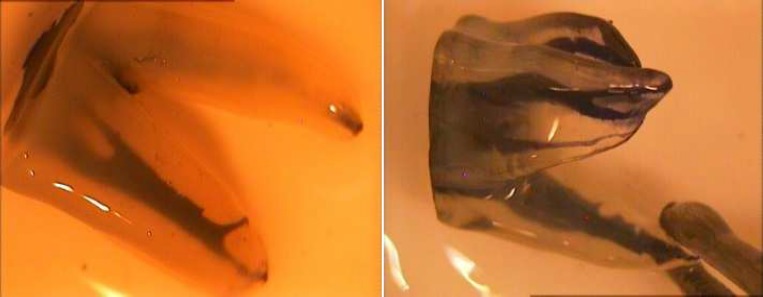
In this study, 80 extracted permanent maxillary first molars from a population in Rafsanjan, Iran were collected. Root canal morphology was evaluated by clearing technique under stereomicroscope under 40× magnification. A combination of Vertucci's and Sert and Bayirli's classifications were used to determine the root canal types. Data were analyzed by SPSS 18 software using descriptive statistics.

RESULTS

All palatal roots and almost all distobuccal roots had type I configuration. Ten different types of root canal system were found in mesiobuccal roots, among which type I was the most common (38.75%), followed by type II, IV, V, VI, IX, XV, XVI=XIX and VII, respectively

CONCLUSION

The mesiobuccal roots of permanent maxillary first molar had the most complex root configuration.

摘要

引言

成功的根管治疗取决于对根管形态及其变异的全面了解、合适的开髓腔、恰当的清理和成形以及充分的根管充填。本研究旨在评估伊朗人群中恒上颌第一磨牙的根管形态。

方法与材料

本研究收集了来自伊朗拉夫桑詹人群的80颗拔除的恒上颌第一磨牙。在体视显微镜下40倍放大倍数下通过透明技术评估根管形态。采用韦尔图奇分类法和塞尔特与巴伊里分类法相结合的方式来确定根管类型。使用SPSS 18软件进行描述性统计分析数据。

结果

所有腭根和几乎所有远中颊根均为I型构型。在近中颊根中发现了10种不同类型的根管系统,其中I型最为常见(38.75%),其次分别为II型、IV型、V型、VI型、IX型、XV型、XVI = XIX型和VII型。

结论

恒上颌第一磨牙的近中颊根具有最复杂的根管形态。

https://cdn.ncbi.nlm.nih.gov/pmc/blobs/0e34/5800446/9d254ce82637/iej-13-078-g001.jpg

相似文献

1
Evaluating the Root Canal Morphology of Permanent Maxillary First Molars in Iranian Population.评估伊朗人群中恒牙上颌第一磨牙的根管形态
Iran Endod J. 2018 Winter;13(1):78-82. doi: 10.22037/iej.v12i4.17207.
2
Evaluation of root canal morphology of maxillary molars using cone beam computed tomography.使用锥形束计算机断层扫描评估上颌磨牙的根管形态
Pak J Med Sci. 2015 Mar-Apr;31(2):426-30. doi: 10.12669/pjms.312.6753.
3
Root canal configuration of permanent maxillary first molar in Khasi population of Meghalaya: An in vitro study.梅加拉亚邦卡西族人群上颌第一恒磨牙的根管形态:一项体外研究。
J Conserv Dent. 2014 Jul;17(4):359-63. doi: 10.4103/0972-0707.136511.
4
Cone-beam computed tomography evaluation of maxillary first and second molars in Iranian population: a morphological study.伊朗人群上颌第一和第二磨牙的锥形束计算机断层扫描评估:一项形态学研究。
Iran Endod J. 2014 Summer;9(3):190-4. Epub 2014 Jul 5.
5
Root Morphology of the Maxillary First and Second Molars in an Iranian Population Using Cone Beam Computed Tomography.使用锥形束计算机断层扫描技术对上颌第一和第二磨牙在伊朗人群中的牙根形态进行研究。
J Dent (Tehran). 2017 May;14(3):115-122.
6
Canal Configuration of Mesiobuccal Roots in Permanent Maxillary First Molars in Iranian Population: A Systematic Review.伊朗人群恒牙上颌第一磨牙近中颊根的根管形态:一项系统评价
J Dent (Tehran). 2016 Nov;13(6):438-447.
7
Root and canal morphology of Thai maxillary molars.泰国上颌磨牙的牙根及根管形态
Int Endod J. 2002 May;35(5):478-85. doi: 10.1046/j.1365-2591.2002.00511.x.
8
A Cone-beam Computed Tomographic Study of Root and Canal Morphology of Maxillary First and Second Permanent Molars in a Thai Population.一项泰国人群上颌第一和第二恒磨牙根管及形态的锥形束 CT 研究。
J Endod. 2018 Jan;44(1):56-61. doi: 10.1016/j.joen.2017.08.020. Epub 2017 Oct 20.
9
Root canal morphology of primary molars by clearing technique: An study.通过透明技术观察乳磨牙的根管形态:一项研究。
J Indian Soc Pedod Prev Dent. 2018 Apr-Jun;36(2):151-157. doi: 10.4103/JISPPD.JISPPD_237_16.
10
Report of a rare case: a maxillary first molar with seven canals confirmed with cone-beam computed tomography.罕见病例报告:经锥形束计算机断层扫描证实的上颌第一磨牙有七条根管。
Iran Endod J. 2014 Spring;9(2):153-7. Epub 2014 Mar 8.

引用本文的文献

1
Prevalence of second mesiobuccal canal in maxillary molars of Iranian population: A systematic review with meta-analysis.伊朗人群上颌磨牙近中颊侧第二根管的患病率:一项Meta分析的系统评价
PLoS One. 2025 Jul 11;20(7):e0327006. doi: 10.1371/journal.pone.0327006. eCollection 2025.
2
Efficacy of Two Different Views of Video Demonstration in Teaching Access Cavity Preparation to Third Year Dental Students.两种不同视角的视频演示在向牙科三年级学生教授备洞操作中的效果。
Iran Endod J. 2018 Fall;13(4):474-480. doi: 10.22037/iej.v13i4.20275.
3
Canal Transportation and Centering after Using PathFile and R-Pilot in Mesiobuccal Canals of Maxillary Molars Using Cone-Beam Computed Tomography.

本文引用的文献

1
Root Canal Morphologies of Mesiobuccal Roots of Maxillary Molars using Cone beam Computed Tomography and Periapical Radiographic Techniques in an Iranian Population.使用锥形束计算机断层扫描和根尖X线摄影技术对伊朗人群上颌磨牙近中颊根根管形态的研究
J Contemp Dent Pract. 2017 Sep 1;18(9):745-749. doi: 10.5005/jp-journals-10024-2119.
2
Root Morphology and Canal Configuration of First and Second Maxillary Molars in a Selected Iranian Population: A Cone-Beam Computed Tomography Evaluation.特定伊朗人群中上颌第一和第二磨牙的牙根形态及根管形态:锥形束计算机断层扫描评估
Iran Endod J. 2017 Summer;12(3):288-292. doi: 10.22037/iej.v12i3.13708.
3
使用锥束计算机断层扫描技术观察PathFile和R-Pilot在磨牙近中颊根管中的管腔交通情况及根管就位情况
Iran Endod J. 2019 Winter;14(1):35-39. doi: 10.22037/iej.v14i1.21601.
4
The MB3 canal in maxillary molars: a micro-CT study.上颌磨牙 MB3 管:一项微 CT 研究。
Clin Oral Investig. 2020 Nov;24(11):4109-4121. doi: 10.1007/s00784-020-03284-7. Epub 2020 May 7.
5
Cone beam computed tomographic survey of the mesiobuccal root canal anatomy in the maxillary first and second molar teeth of an Iranian population.伊朗人群上颌第一和第二磨牙近中颊根管解剖结构的锥形束计算机断层扫描研究。
Eur J Dent. 2018 Jul-Sep;12(3):422-427. doi: 10.4103/ejd.ejd_60_18.
Canal Configuration of Mesiobuccal Roots in Permanent Maxillary First Molars in Iranian Population: A Systematic Review.
伊朗人群恒牙上颌第一磨牙近中颊根的根管形态:一项系统评价
J Dent (Tehran). 2016 Nov;13(6):438-447.
4
Root Canal Anatomy and Morphology of Mandibular First Molars in a Selected Iranian Population: An Study.特定伊朗人群下颌第一磨牙的根管解剖与形态:一项研究
Iran Endod J. 2017 Winter;12(1):87-91. doi: 10.22037/iej.2017.18.
5
Root Canal Morphology of Mandibular Canine in an Iranian Population: A CBCT Assessment.伊朗人群下颌尖牙的根管形态:CBCT评估
Iran Endod J. 2017 Winter;12(1):78-82. doi: 10.22037/iej.2017.16.
6
A Review on Root Anatomy and Canal Configuration of the Maxillary Second Molars.上颌第二磨牙的牙根解剖与根管形态综述
Iran Endod J. 2017 Winter;12(1):1-9. doi: 10.22037/iej.2017.01.
7
The efficacy of sulfated polysaccharides from Padina tetrastromatica in modulating the immune functions of RAW 264.7 cells.四列藻硫酸化多糖对RAW 264.7细胞免疫功能的调节作用
Biomed Pharmacother. 2017 Apr;88:677-683. doi: 10.1016/j.biopha.2017.01.094. Epub 2017 Jan 30.
8
Survey of Anatomy and Root Canal Morphology of Maxillary First Molars Regarding Age and Gender in an Iranian Population Using Cone-Beam Computed Tomography.使用锥形束计算机断层扫描对伊朗人群中上颌第一磨牙的解剖结构和根管形态进行年龄和性别的调查。
Iran Endod J. 2016 Fall;11(4):298-303. doi: 10.22037/iej.2016.8.
9
A Clinical Update on the Different Methods to Decrease the Occurrence of Missed Root Canals.减少遗漏根管发生的不同方法的临床最新进展
Iran Endod J. 2016 Summer;11(3):208-13. doi: 10.7508/iej.2016.03.012. Epub 2016 May 1.
10
Cone-beam computed tomography evaluation of maxillary first and second molars in Iranian population: a morphological study.伊朗人群上颌第一和第二磨牙的锥形束计算机断层扫描评估:一项形态学研究。
Iran Endod J. 2014 Summer;9(3):190-4. Epub 2014 Jul 5.