Hajizadeh Niusha, Madani Zahra Sadat, Zabihi Ebrahim, Golpour Moniyreh, Zahedpasha Amir, Mohammadnia Mousa
Department of Endodontics, School of Dentistry, Babol University of Medical Sciences, Babol, Iran.
Dental Materials Research Center, School of Dentistry, Babol University of Medical Sciences, Babol, Iran.
Iran Endod J. 2018 Winter;13(1):94-101. doi: 10.22037/iej.v13i1.17860.
This study assessed the effect of mineral trioxide aggregate (MTA) and calcium-enriched mixture (CEM) cement on odontogenic differentiation and mineralization of stem cells.
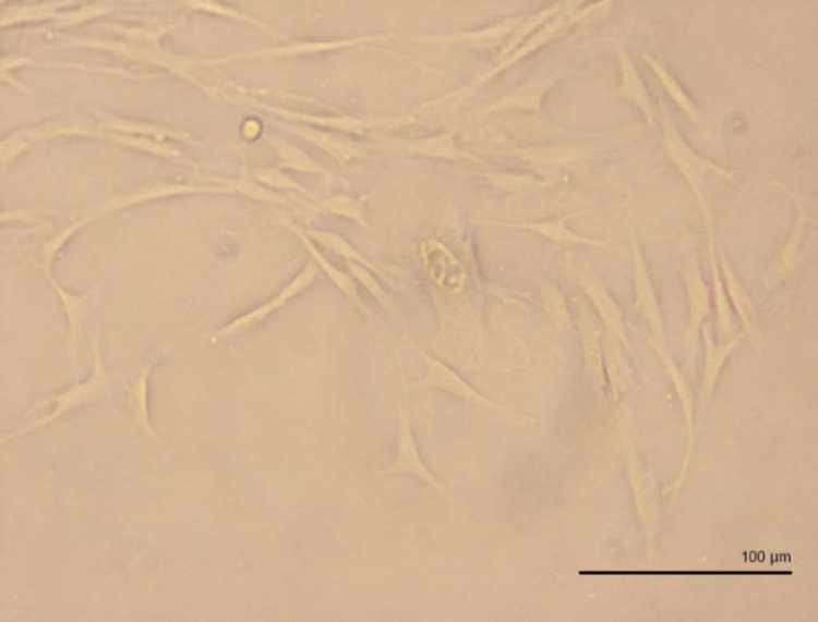
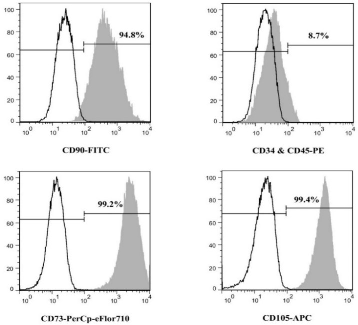
After confirmation of stemness and homogeneity of stem cells derived from apical papilla (SCAPs) using flow cytometry, the cells were exposed for 3 weeks to either osteogenic medium (OS) or CEM extract+OS (CEM+OS) or MTA extract in OS (MTA+OS) or DMEM based regular culture media (negative control). Relative expression of alkaline phosphatase (ALP), dentine sialophosphoprotein (DSPP), osteocalcin (OSC), and osterix (SP7) were measured at days 14 and 21 using RT-qPCR method. At the same time points Alizarin Red staining method was used to assess mineralization potential of SCAPS. Gene expression changes analysis were made automatically using REST® software and a <0.05 was considered significant.
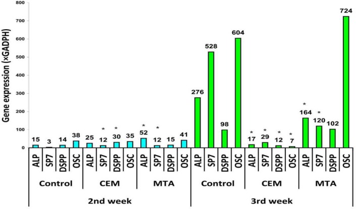
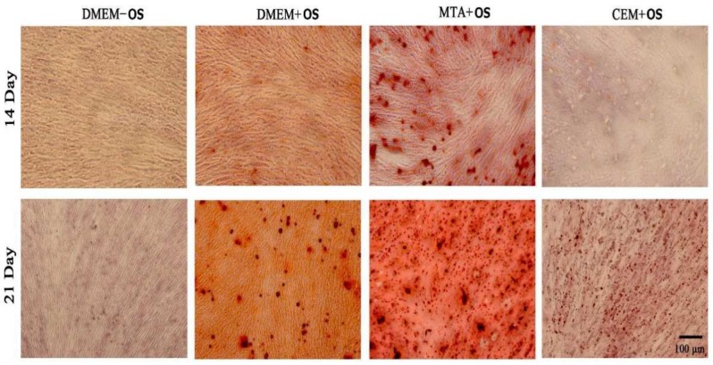
After 2 weeks of exposure, expression of all genes were between 3 and 52 times the expression of GADPH (all were upregulated except SP7 in the control, <0.05). After 3 weeks, relative expressions of the genes: ALP, SP7, DSPP, and OSC were respectively 275.9, 528.3, 98.4, and 603.7 times the expression of GADPH in the control group (OS). These were respectively 17.405, 29.2, 11.8, and 6.5 in CEM+OS group, and 163.8, 119.7, 102.5, and 723.9 in MTA+OS group. All of these were confirmed as upregulated (<0.05) except for ALP and OSC of DM+CEM group. After 2 weeks, alizarin red staining showed similar mineralized nodules in OS, MTA+OS, and CEM+OS. In third week, larger nodules were seen in MTA+OS and OS, but not in CEM+OS.
After 2 weeks, gene expressions were almost comparable in OS, CEM+OS, and MTA+OS. After 3 weeks, OS and MTA+OS upregulated genes much greater than in 2nd week. However, upregulation in CEM+OS might not increase in 3rd week compared to those in 2nd week.
本研究评估了三氧化矿物凝聚体(MTA)和富钙混合物(CEM)水泥对干细胞牙源性分化和矿化的影响。
使用流式细胞术确认根尖乳头干细胞(SCAPs)的干性和同质性后,将细胞在成骨培养基(OS)或CEM提取物+OS(CEM+OS)或OS中的MTA提取物(MTA+OS)或基于DMEM的常规培养基(阴性对照)中培养3周。在第14天和第21天使用RT-qPCR方法测量碱性磷酸酶(ALP)、牙本质涎磷蛋白(DSPP)、骨钙素(OSC)和osterix(SP7)的相对表达。在相同时间点,使用茜素红染色法评估SCAPs的矿化潜力。使用REST®软件自动进行基因表达变化分析,P<0.05被认为具有统计学意义。
暴露2周后,所有基因的表达是GADPH表达的3至52倍(对照组中除SP7外均上调,P<0.05)。3周后,基因ALP、SP7、DSPP和OSC的相对表达分别是对照组(OS)中GADPH表达的275.9、528.3、98.4和603.7倍。在CEM+OS组中分别为17.405、29.2、11.8和6.5,在MTA+OS组中分别为163.8、119.7、102.5和723.9。除DM+CEM组的ALP和OSC外,所有这些均被确认为上调(P<0.05)。2周后,茜素红染色显示OS、MTA+OS和CEM+OS中有相似的矿化结节。在第3周,MTA+OS和OS中可见较大的结节,但CEM+OS中未见。
2周后,OS、CEM+OS和MTA+OS中的基因表达几乎相当。3周后,OS和MTA+OS上调的基因比第2周大得多。然而与第2周相比,CEM+OS在第3周的上调可能没有增加。