• 文献检索
  • 文档翻译
  • 深度研究
  • 学术资讯
  • Suppr Zotero 插件Zotero 插件
  • 邀请有礼
  • 套餐&价格
  • 历史记录
应用&插件
Suppr Zotero 插件Zotero 插件浏览器插件Mac 客户端Windows 客户端微信小程序
定价
高级版会员购买积分包购买API积分包
服务
文献检索文档翻译深度研究API 文档MCP 服务
关于我们
关于 Suppr公司介绍联系我们用户协议隐私条款
关注我们

Suppr 超能文献

核心技术专利:CN118964589B侵权必究
粤ICP备2023148730 号-1Suppr @ 2026

文献检索

告别复杂PubMed语法,用中文像聊天一样搜索,搜遍4000万医学文献。AI智能推荐,让科研检索更轻松。

立即免费搜索

文件翻译

保留排版,准确专业,支持PDF/Word/PPT等文件格式,支持 12+语言互译。

免费翻译文档

深度研究

AI帮你快速写综述,25分钟生成高质量综述,智能提取关键信息,辅助科研写作。

立即免费体验

对比增强灌注磁共振成像鉴别复发性/残留性脑肿瘤与放射性坏死

Contrast-Enhanced Perfusion MR Imaging to Differentiate Between Recurrent/Residual Brain Neoplasms and Radiation Necrosis.

作者信息

Metaweh Noha Ahmed Kamal, Azab Amr Osama, El Basmy Ayman Abd El Hameed, Mashhour Karim Nabil, El Mahdy Wael Mokhtar

机构信息

Radiology Department, Faculty of Medicine, Cairo University, Cairo, Egypt. Email:

出版信息

Asian Pac J Cancer Prev. 2018 Apr 25;19(4):941-948. doi: 10.22034/APJCP.2018.19.4.941.

DOI:10.22034/APJCP.2018.19.4.941
PMID:29693348
原文链接:https://pmc.ncbi.nlm.nih.gov/articles/PMC6031785/
Abstract

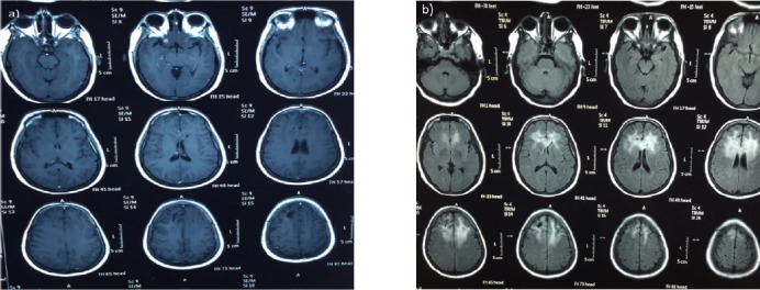
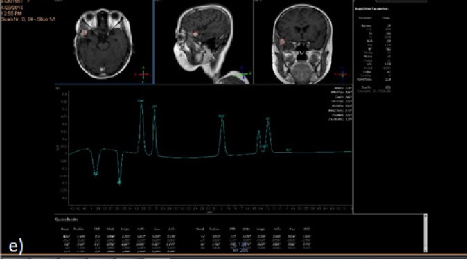
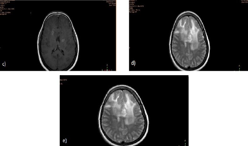
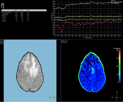
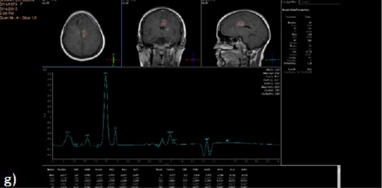
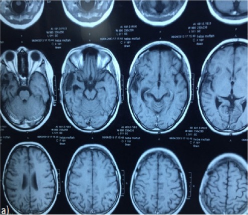
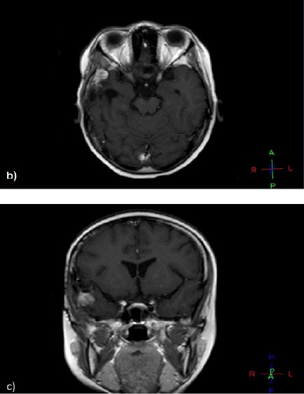
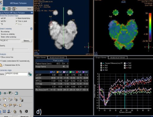
Purpose: To determine the value of dynamic susceptibility contrast enhanced (DSC) MRI (magnetic resonance imaging) perfusion in the characterization of newly developed/enlarging lesions within irradiated regions after treatment of brain tumors. Methods: This prospective cross-sectional study covered 23 patients, 12 females and 11 males. All cases initially presented with histologically proven malignant brain tumors and underwent surgical intervention followed by radiotherapy (+/- chemotherapy). On follow up imaging, they presented with newly developed/progressively enhancing mass lesions at the sites of the primary tumors. All patients then underwent conventional MRI, DSC MRI perfusion and MR spectroscopy. Results: In our study, we found DSC MR perfusion to be a useful non-invasive method for differentiating recurrent brain tumors from radiation necrosis. This approach allows hemodynamic measurements to be obtained within the brain as the relative cerebral blood volume (rCBV) to complement the anatomic information obtained with conventional contrast enhanced MR imaging. The sensitivity and specificity of DSC MR perfusion for differentiation were found to be 77.8% and 80.0%, respectively. Conclusion: DSC MR perfusion is a promising technique in differentiating recurrent brain tumors from radiation necrosis as it has acceptable spatial resolution and can be routinely performed in the same settings after conventional MRI.

摘要

目的

确定动态磁敏感对比增强(DSC)磁共振成像(MRI)灌注在脑肿瘤治疗后照射区域内新出现/增大病变特征分析中的价值。方法:这项前瞻性横断面研究涵盖了23例患者,其中女性12例,男性11例。所有病例最初均经组织学证实为恶性脑肿瘤,并接受了手术干预,随后进行放疗(±化疗)。在随访成像中,他们在原发肿瘤部位出现了新出现/逐渐增强的肿块病变。所有患者随后均接受了常规MRI、DSC MRI灌注和磁共振波谱检查。结果:在我们的研究中,我们发现DSC MR灌注是一种用于区分复发性脑肿瘤与放射性坏死的有用的非侵入性方法。这种方法能够在脑内获得血流动力学测量值,即相对脑血容量(rCBV),以补充通过常规对比增强MR成像获得的解剖学信息。DSC MR灌注用于鉴别的敏感性和特异性分别为77.8%和80.0%。结论:DSC MR灌注在区分复发性脑肿瘤与放射性坏死方面是一种有前景的技术,因为它具有可接受的空间分辨率,并且可以在常规MRI后的相同条件下常规进行。

https://cdn.ncbi.nlm.nih.gov/pmc/blobs/d60d/6031785/209252877a56/APJCP-19-941-g008.jpg
https://cdn.ncbi.nlm.nih.gov/pmc/blobs/d60d/6031785/d36692f98fb8/APJCP-19-941-g001.jpg
https://cdn.ncbi.nlm.nih.gov/pmc/blobs/d60d/6031785/fde38f591835/APJCP-19-941-g002.jpg
https://cdn.ncbi.nlm.nih.gov/pmc/blobs/d60d/6031785/9a58aba50b78/APJCP-19-941-g003.jpg
https://cdn.ncbi.nlm.nih.gov/pmc/blobs/d60d/6031785/38a177337e88/APJCP-19-941-g004.jpg
https://cdn.ncbi.nlm.nih.gov/pmc/blobs/d60d/6031785/71225e6011cb/APJCP-19-941-g005.jpg
https://cdn.ncbi.nlm.nih.gov/pmc/blobs/d60d/6031785/93085e5cc672/APJCP-19-941-g006.jpg
https://cdn.ncbi.nlm.nih.gov/pmc/blobs/d60d/6031785/e1069088c6b0/APJCP-19-941-g007.jpg
https://cdn.ncbi.nlm.nih.gov/pmc/blobs/d60d/6031785/209252877a56/APJCP-19-941-g008.jpg

相似文献

1
Contrast-Enhanced Perfusion MR Imaging to Differentiate Between Recurrent/Residual Brain Neoplasms and Radiation Necrosis.对比增强灌注磁共振成像鉴别复发性/残留性脑肿瘤与放射性坏死
Asian Pac J Cancer Prev. 2018 Apr 25;19(4):941-948. doi: 10.22034/APJCP.2018.19.4.941.
2
Comparison of Diffusion Tensor Imaging and Magnetic Resonance Perfusion Imaging in Differentiating Recurrent Brain Neoplasm From Radiation Necrosis.扩散张量成像与磁共振灌注成像在鉴别复发性脑肿瘤与放射性坏死中的比较
Acad Radiol. 2016 May;23(5):569-76. doi: 10.1016/j.acra.2015.11.015. Epub 2016 Feb 23.
3
Differentiation of recurrent glioblastoma multiforme from radiation necrosis after external beam radiation therapy with dynamic susceptibility-weighted contrast-enhanced perfusion MR imaging.采用动态磁敏感加权对比增强灌注磁共振成像鉴别多形性胶质母细胞瘤复发与外照射放疗后放射性坏死
Radiology. 2009 Nov;253(2):486-96. doi: 10.1148/radiol.2532090007. Epub 2009 Sep 29.
4
Dynamic susceptibility contrast (DSC) perfusion MRI in differential diagnosis between radionecrosis and neoangiogenesis in cerebral metastases using rCBV, rCBF and K2.动态对比磁共振灌注成像(DSC)在脑转移瘤放射性坏死与新生血管形成鉴别诊断中的应用:rCBV、rCBF 和 K2 的价值。
Radiol Med. 2018 Jul;123(7):545-552. doi: 10.1007/s11547-018-0866-7. Epub 2018 Mar 5.
5
Perfusion weighted magnetic resonance imaging to distinguish the recurrence of metastatic brain tumors from radiation necrosis after stereotactic radiosurgery.磁共振灌注成像鉴别立体定向放射外科治疗后脑转移瘤复发与放射性坏死
J Neurooncol. 2010 Aug;99(1):81-8. doi: 10.1007/s11060-009-0106-z. Epub 2010 Jan 8.
6
Posttherapeutic intraaxial brain tumor: the value of perfusion-sensitive contrast-enhanced MR imaging for differentiating tumor recurrence from nonneoplastic contrast-enhancing tissue.治疗后轴内脑肿瘤:灌注敏感对比增强磁共振成像在鉴别肿瘤复发与非肿瘤性对比增强组织中的价值。
AJNR Am J Neuroradiol. 2000 May;21(5):901-9.
7
DCE and DSC perfusion MRI diagnostic accuracy in the follow-up of primary and metastatic intra-axial brain tumors treated by radiosurgery with cyberknife.DCE 和 DSC 灌注 MRI 在接受 Cyberknife 放射外科治疗的原发性和转移性脑内肿瘤随访中的诊断准确性。
Radiat Oncol. 2019 Apr 15;14(1):65. doi: 10.1186/s13014-019-1271-7.
8
Comparison of three different MR perfusion techniques and MR spectroscopy for multiparametric assessment in distinguishing recurrent high-grade gliomas from stable disease.比较三种不同的磁共振灌注技术和磁共振波谱在鉴别复发性高级别胶质瘤与稳定期疾病中的多参数评估。
Acad Radiol. 2013 Dec;20(12):1557-65. doi: 10.1016/j.acra.2013.09.003.
9
Differentiating Radiation-Induced Necrosis from Recurrent Brain Tumor Using MR Perfusion and Spectroscopy: A Meta-Analysis.使用磁共振灌注成像和波谱分析鉴别放射性坏死与复发性脑肿瘤:一项荟萃分析
PLoS One. 2016 Jan 7;11(1):e0141438. doi: 10.1371/journal.pone.0141438. eCollection 2016.
10
Differentiation between radiation-induced brain injury and glioma recurrence using 3D pCASL and dynamic susceptibility contrast-enhanced perfusion-weighted imaging.采用 3D pCASL 和动态磁敏感对比增强灌注加权成像鉴别放射性脑损伤与胶质瘤复发。
Radiother Oncol. 2018 Oct;129(1):68-74. doi: 10.1016/j.radonc.2018.01.009. Epub 2018 Feb 2.

引用本文的文献

1
Comparison of Imaging Modalities in Differentiating Cerebral Neoplastic Lesions and Post-radiation Necrosis.鉴别脑肿瘤性病变与放射性坏死的成像模态比较
Cureus. 2025 Feb 6;17(2):e78653. doi: 10.7759/cureus.78653. eCollection 2025 Feb.
2
Evaluating the Efficacy of Perfusion MRI and Conventional MRI in Distinguishing Recurrent Cerebral Metastasis from Brain Radiation Necrosis.评估灌注磁共振成像和传统磁共振成像在鉴别复发性脑转移瘤与脑放射性坏死中的效能。
Brain Sci. 2024 Mar 27;14(4):321. doi: 10.3390/brainsci14040321.
3
Radionecrosis mimicking pseudo‑progression in a patient with lung cancer and brain metastasis following the combination of anti‑PD‑1 therapy and stereotactic radiosurgery: A case report.

本文引用的文献

1
Differentiating Radiation-Induced Necrosis from Recurrent Brain Tumor Using MR Perfusion and Spectroscopy: A Meta-Analysis.使用磁共振灌注成像和波谱分析鉴别放射性坏死与复发性脑肿瘤:一项荟萃分析
PLoS One. 2016 Jan 7;11(1):e0141438. doi: 10.1371/journal.pone.0141438. eCollection 2016.
2
Glioblastoma treated with concurrent radiation therapy and temozolomide chemotherapy: differentiation of true progression from pseudoprogression with quantitative dynamic contrast-enhanced MR imaging.胶质母细胞瘤行同期放化疗治疗:定量动态对比增强磁共振成像鉴别真性进展与假性进展。
Radiology. 2015 Mar;274(3):830-40. doi: 10.1148/radiol.14132632. Epub 2014 Oct 21.
3
抗PD-1治疗联合立体定向放射外科治疗后肺癌脑转移患者中类似假性进展的放射性坏死:一例报告
Oncol Lett. 2023 Jul 6;26(2):361. doi: 10.3892/ol.2023.13947. eCollection 2023 Aug.
4
Distinguishing Tumor Admixed in a Radiation Necrosis (RN) Background: H and H MR With a Novel Mouse Brain-Tumor/RN Model.鉴别混杂于放射性坏死(RN)背景中的肿瘤:采用新型小鼠脑肿瘤/RN模型的氢质子磁共振波谱分析(H and H MR)
Front Oncol. 2022 May 30;12:885480. doi: 10.3389/fonc.2022.885480. eCollection 2022.
5
Osteoradionecrosis of the craniotomy flap: a rare complication of stereotactic radiosurgery.开颅皮瓣放射性骨坏死:立体定向放射外科的一种罕见并发症。
Oxf Med Case Reports. 2022 Apr 19;2022(4):omac032. doi: 10.1093/omcr/omac032. eCollection 2022 Apr.
6
Radiomics-Based Detection of Radionecrosis Using Harmonized Multiparametric MRI.基于影像组学的放射性坏死的多参数MRI检测
Cancers (Basel). 2022 Jan 7;14(2):286. doi: 10.3390/cancers14020286.
7
Tumor recurrence or treatment-related changes following chemoradiation in patients with glioblastoma: does pathology predict outcomes?胶质母细胞瘤患者放化疗后肿瘤复发或治疗相关改变:病理学能否预测结局?
J Neurooncol. 2021 Mar;152(1):163-172. doi: 10.1007/s11060-020-03690-7. Epub 2021 Jan 22.
Which combination of MR imaging modalities is best for predicting recurrent glioblastoma? Study of diagnostic accuracy and reproducibility.
哪种磁共振成像方式组合最适合预测复发性脑胶质瘤?诊断准确性和可重复性研究。
Radiology. 2014 Dec;273(3):831-43. doi: 10.1148/radiol.14132868. Epub 2014 May 30.
4
Pseudoprogression of glioblastoma after chemo- and radiation therapy: diagnosis by using dynamic susceptibility-weighted contrast-enhanced perfusion MR imaging with ferumoxytol versus gadoteridol and correlation with survival.替莫唑胺化疗和放疗后胶质母细胞瘤的假性进展:使用氧化铁对比剂 ferumoxytol 与钆喷酸葡胺进行动态磁敏感对比增强灌注 MRI 诊断与生存相关性研究
Radiology. 2013 Mar;266(3):842-52. doi: 10.1148/radiol.12111472. Epub 2012 Nov 30.
5
Reevaluating the imaging definition of tumor progression: perfusion MRI quantifies recurrent glioblastoma tumor fraction, pseudoprogression, and radiation necrosis to predict survival.重新评估肿瘤进展的影像学定义:灌注 MRI 定量评估复发性脑胶质瘤肿瘤部分、假性进展和放射性坏死,以预测患者的生存情况。
Neuro Oncol. 2012 Jul;14(7):919-30. doi: 10.1093/neuonc/nos112. Epub 2012 May 3.
6
Treatment-related change versus tumor recurrence in high-grade gliomas: a diagnostic conundrum--use of dynamic susceptibility contrast-enhanced (DSC) perfusion MRI.高级别胶质瘤中的治疗相关性变化与肿瘤复发:诊断难题——动态磁敏感对比增强(DSC)灌注 MRI 的应用。
AJR Am J Roentgenol. 2012 Jan;198(1):19-26. doi: 10.2214/AJR.11.7417.
7
Posttreatment high-grade glioma: usefulness of peak height position with semiquantitative MR perfusion histogram analysis in an entire contrast-enhanced lesion for predicting volume fraction of recurrence.治疗后高级别胶质瘤:半定量 MR 灌注直方图分析整个增强病变的峰值高度位置在预测复发体积分数中的作用。
Radiology. 2010 Sep;256(3):906-15. doi: 10.1148/radiol.10091461. Epub 2010 Jul 15.
8
Differentiation between brain tumor recurrence and radiation injury using perfusion, diffusion-weighted imaging and MR spectroscopy.使用灌注、弥散加权成像和磁共振波谱区分脑肿瘤复发与放射性损伤。
Folia Neuropathol. 2010;48(2):81-92.
9
Relative value of magnetic resonance spectroscopy, magnetic resonance perfusion, and 2-(18F) fluoro-2-deoxy-D-glucose positron emission tomography for detection of recurrence or grade increase in gliomas.磁共振波谱、磁共振灌注和 2-(18F)氟-2-脱氧-D-葡萄糖正电子发射断层扫描在检测胶质瘤复发或分级增加中的相对价值。
J Clin Neurosci. 2010 Jan;17(1):50-3. doi: 10.1016/j.jocn.2009.02.035. Epub 2009 Dec 2.
10
Distinction between glioma progression and post-radiation change by combined physiologic MR imaging.结合生理 MRI 鉴别脑胶质瘤进展与放疗后改变。
Neuroradiology. 2010 Apr;52(4):297-306. doi: 10.1007/s00234-009-0613-9. Epub 2009 Oct 16.