Program in Biophysics, Stanford University, Stanford, CA, USA.
Department of Structural Biology, Stanford University School of Medicine, Stanford, CA, USA.
Nature. 2018 May;557(7703):118-122. doi: 10.1038/s41586-018-0055-9. Epub 2018 Apr 25.
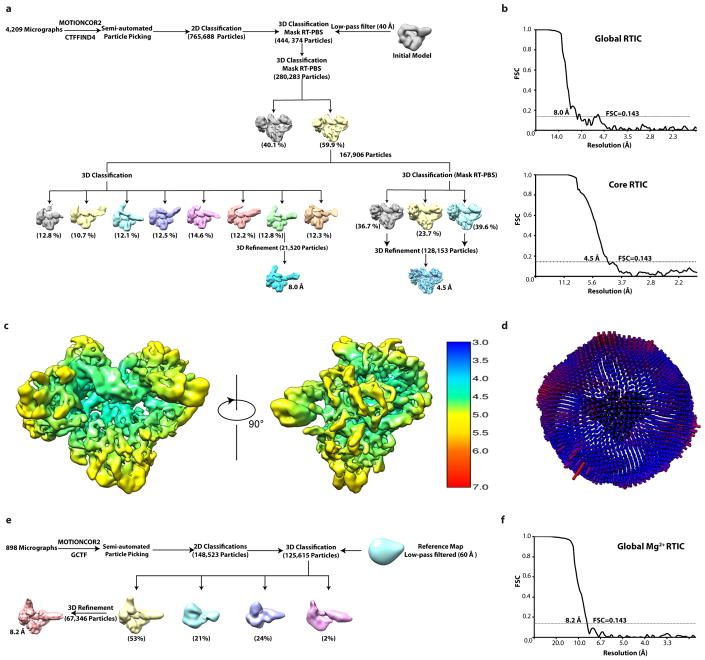
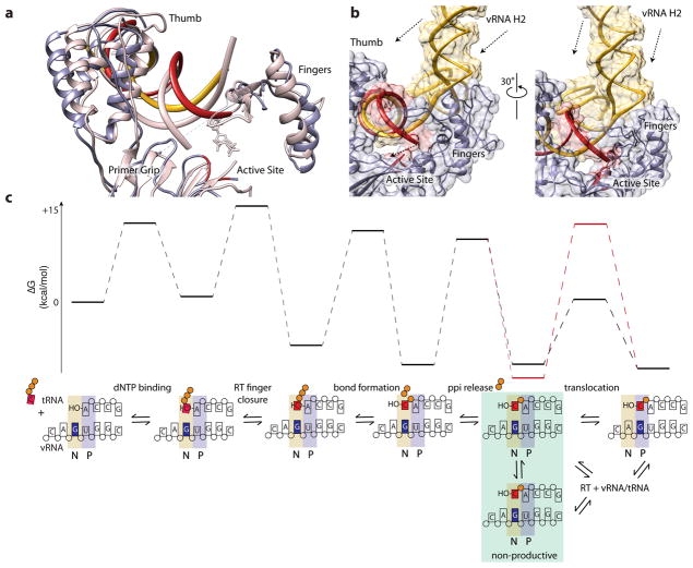
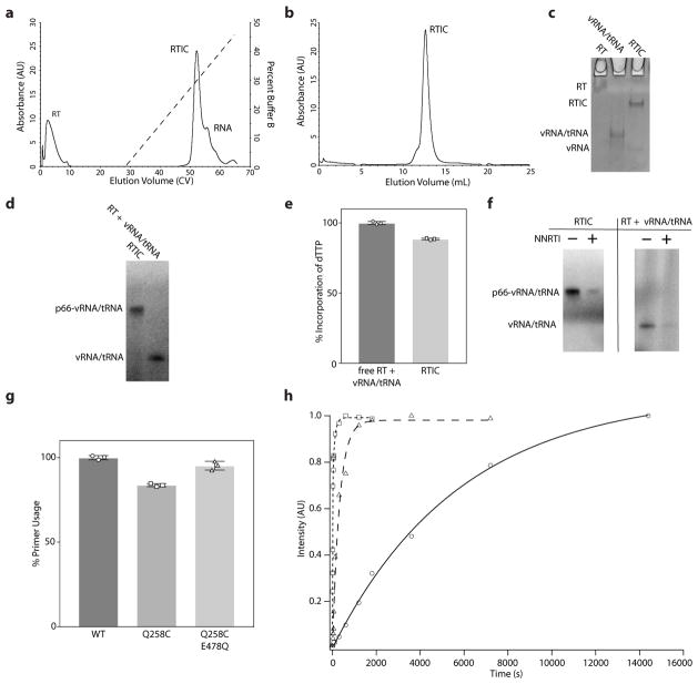
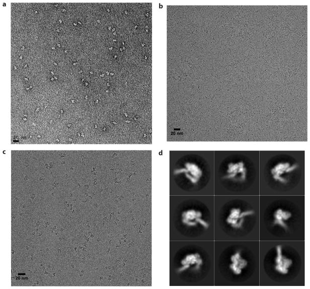
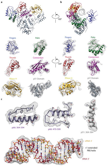
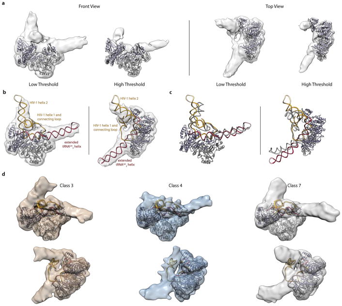
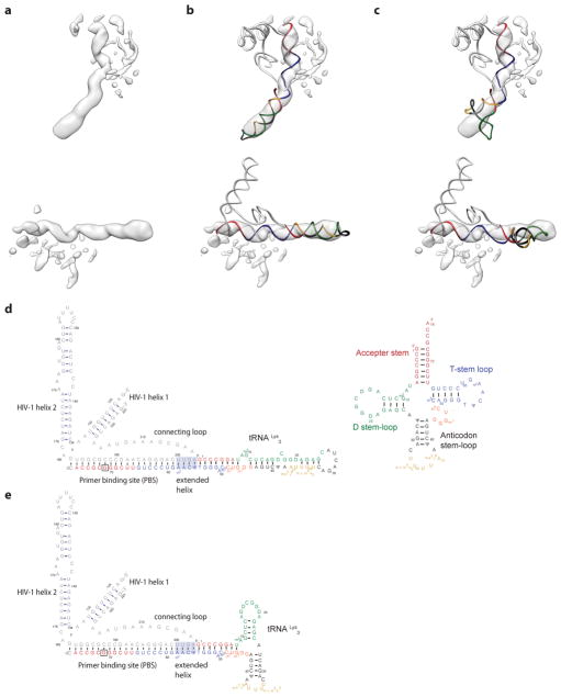
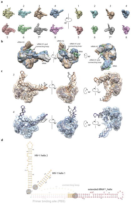
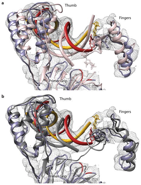
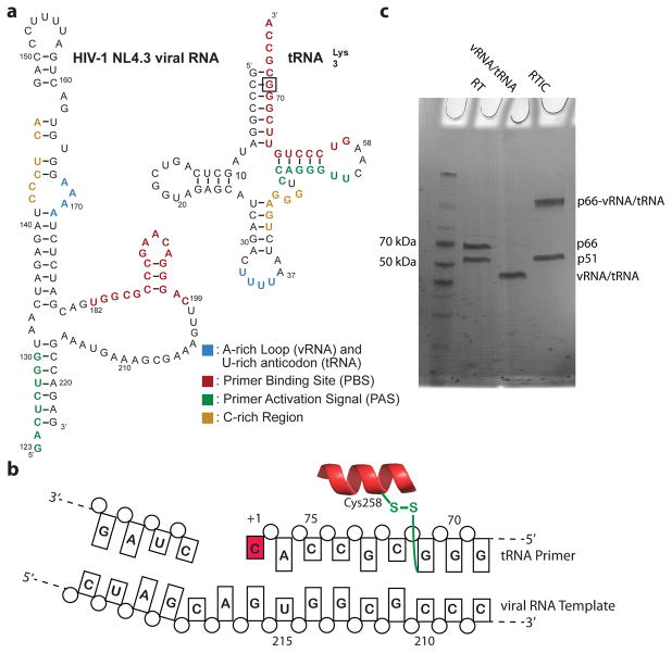
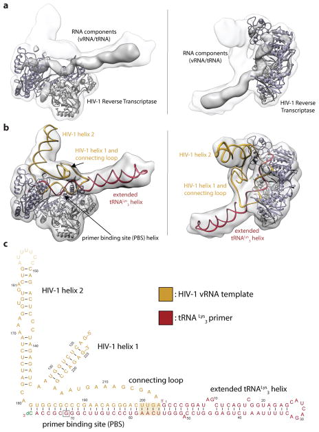
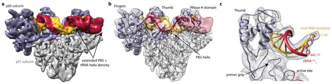
Reverse transcription of the HIV-1 RNA genome into double-stranded DNA is a central step in viral infection and a common target of antiretroviral drugs . The reaction is catalysed by viral reverse transcriptase (RT) that is packaged in an infectious virion with two copies of viral genomic RNA each bound to host lysine 3 transfer RNA (tRNA), which acts as a primer for initiation of reverse transcription. Upon viral entry into cells, initiation is slow and non-processive compared to elongation. Despite extensive efforts, the structural basis of RT function during initiation has remained a mystery. Here we use cryo-electron microscopy to determine a three-dimensional structure of an HIV-1 RT initiation complex. In our structure, RT is in an inactive polymerase conformation with open fingers and thumb and with the nucleic acid primer-template complex shifted away from the active site. The primer binding site (PBS) helix formed between tRNA and HIV-1 RNA lies in the cleft of RT and is extended by additional pairing interactions. The 5' end of the tRNA refolds and stacks on the PBS to create a long helical structure, while the remaining viral RNA forms two helical stems positioned above the RT active site, with a linker that connects these helices to the RNase H region of the PBS. Our results illustrate how RNA structure in the initiation complex alters RT conformation to decrease activity, highlighting a potential target for drug action.
HIV-1 RNA 基因组的逆转录成双链 DNA 是病毒感染的一个关键步骤,也是抗逆转录病毒药物的常见靶点。该反应由病毒逆转录酶 (RT) 催化,RT 与病毒基因组 RNA 的两个拷贝包装在一起,每个拷贝都与宿主赖氨酸 3 转移 RNA (tRNA) 结合,tRNA 作为逆转录起始的引物。病毒进入细胞后,与延伸相比,起始过程缓慢且非连续性。尽管进行了广泛的研究,但 RT 在起始过程中的功能的结构基础仍然是一个谜。在这里,我们使用冷冻电子显微镜来确定 HIV-1 RT 起始复合物的三维结构。在我们的结构中,RT 处于无活性聚合酶构象,其张开的手指和拇指以及与活性位点分离的核酸引物-模板复合物。在 tRNA 和 HIV-1 RNA 之间形成的引物结合位点 (PBS) 螺旋位于 RT 的裂隙中,并通过额外的配对相互作用延伸。tRNA 的 5' 端折叠并堆积在 PBS 上,形成一个长的螺旋结构,而其余的病毒 RNA 形成两个位于 RT 活性位点上方的螺旋茎,连接这些螺旋的链接与 PBS 的 RNase H 区域相连。我们的结果说明了起始复合物中的 RNA 结构如何改变 RT 构象以降低活性,突出了药物作用的潜在靶点。