Department of Surgical Oncology, Osaka City University Graduate School of Medicine, Abeno-ku, Osaka, Japan.
PLoS One. 2018 Apr 26;13(4):e0192744. doi: 10.1371/journal.pone.0192744. eCollection 2018.
Numerous reports indicate that tumor-infiltrating lymphocytes (TILs) are a prognostic factor in various cancers and that they must be good biomarkers. However, the methods of evaluating TILs differ in each study; thus, there is not yet a standardized methodology for evaluating TILs. The purpose of this study is to evaluate the prognostic significance of tumor-infiltrating lymphocytes (TILs) in patients with colorectal cancer (CRC) using the new method proposed by the International TILs Working Group in breast cancer and to standardize the method of evaluating TILs in CRC.
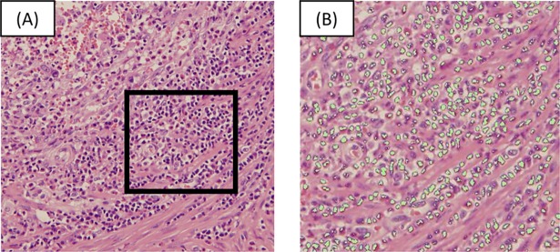
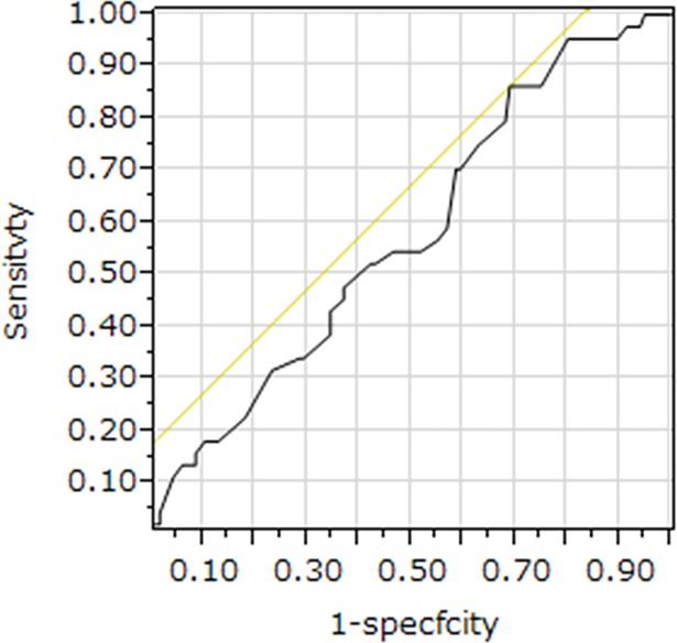
We retrospectively reviewed a database of 160 patients with Stage II or III CRC. The density of TILs was assessed by measuring the area occupied by mononuclear cells over the stromal area on hematoxylin and eosin (H-E)-stained sections. We set 42% as the cut-off percentage of the area occupied by TILs according to the receiver operating characteristic curve, and we classified patients into the high-TILs and the low-TILs groups.
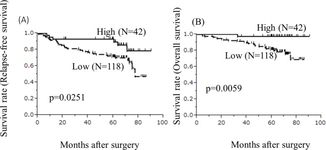
The rates of relapse-free survival (RFS) and overall survival (OS) in the high-TILs group were significantly higher than those in the low-TILs group. A multivariate analysis showed that the density of TILs was independently associated with RFS and OS. Moreover, the density of TILs assessed by an observer was significantly associated with the density of TILs assessed by the automated imaging software program.
The new method for evaluating TILs, which was recommended by the International TILs Working Group in breast cancer, might be a useful predictive factor in colorectal cancer patients.
大量报告表明肿瘤浸润淋巴细胞(TILs)是各种癌症的预后因素,并且它们必须是良好的生物标志物。然而,每个研究中评估 TILs 的方法都不同;因此,目前还没有评估 TILs 的标准化方法。本研究旨在使用国际乳腺癌 TILs 工作组提出的新方法评估结直肠癌(CRC)患者的肿瘤浸润淋巴细胞(TILs)的预后意义,并标准化 CRC 中 TILs 的评估方法。
我们回顾性分析了 160 例 II 期或 III 期 CRC 患者的数据库。通过测量苏木精和伊红(H-E)染色切片上单核细胞占据的区域与基质区域的比例来评估 TILs 的密度。我们根据受试者工作特征曲线将 TILs 占据面积的 42%设定为截止百分比,并将患者分为高 TILs 组和低 TILs 组。
高 TILs 组的无复发生存率(RFS)和总生存率(OS)明显高于低 TILs 组。多变量分析表明,TILs 的密度与 RFS 和 OS 独立相关。此外,观察者评估的 TILs 密度与自动化成像软件程序评估的 TILs 密度显著相关。
国际乳腺癌 TILs 工作组推荐的评估 TILs 的新方法可能是结直肠癌患者的有用预测因素。