Department of Radiology, Charité - Universitätsmedizin Berlin, corporate member of Freie Universität Berlin, Humboldt-Universität zu Berlin, and Berlin Institute of Health, Berlin, Germany.
Richard and Loan Hill Department of Bioengineering, College of Medicine and College of Engineering, University of Illinois at Chicago, Chicago, Illinois, United States of America.
PLoS One. 2018 Apr 26;13(4):e0196486. doi: 10.1371/journal.pone.0196486. eCollection 2018.
Although it has been known for decades that patients with alpha1-antitrypsin deficiency (AATD) have an increased risk of cirrhosis and hepatocellular carcinoma, limited data exist on non-invasive imaging-based methods for assessing liver fibrosis such as magnetic resonance elastography (MRE) and acoustic radiation force impulse (ARFI) quantification, and no data exist on 2D-shear wave elastography (2D-SWE). Therefore, the purpose of this study is to evaluate and compare the applicability of different elastography methods for the assessment of AATD-related liver fibrosis.
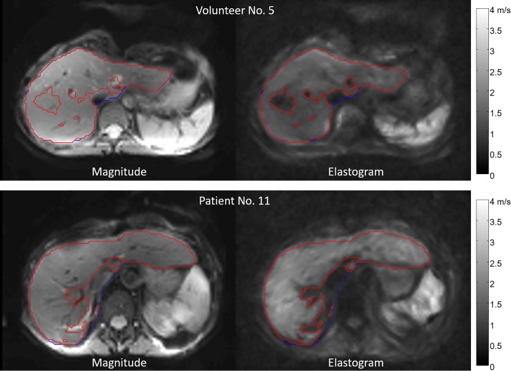
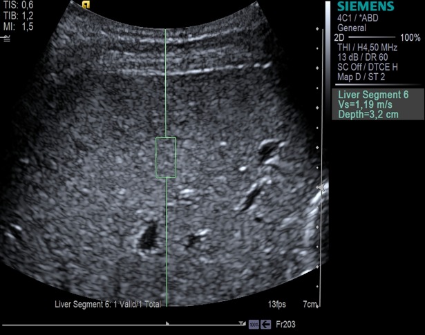
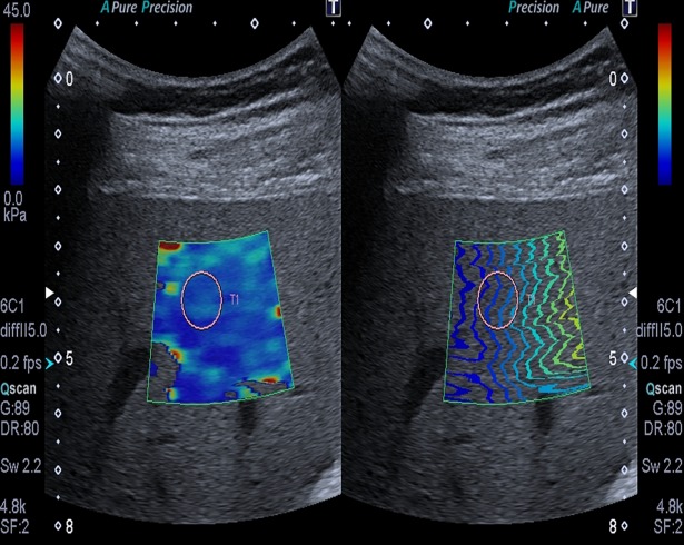
Fifteen clinically asymptomatic AATD patients (11 homozygous PiZZ, 4 heterozygous PiMZ) and 16 matched healthy volunteers were examined using MRE and ARFI quantification. Additionally, patients were examined with 2D-SWE.
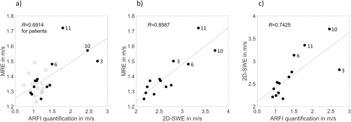
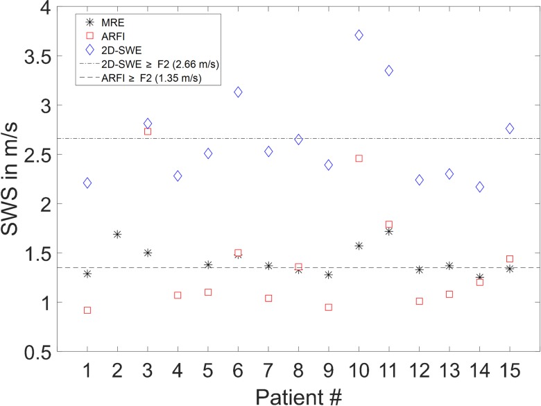
A high correlation is evident for the shear wave speed (SWS) determined with different elastography methods in AATD patients: 2D-SWE/MRE, ARFI quantification/2D-SWE, and ARFI quantification/MRE (R = 0.8587, 0.7425, and 0.6914, respectively; P≤0.0089). Four AATD patients with pathologically increased SWS were consistently identified with all three methods-MRE, ARFI quantification, and 2D-SWE.
The high correlation and consistent identification of patients with pathologically increased SWS using MRE, ARFI quantification, and 2D-SWE suggest that elastography has the potential to become a suitable imaging tool for the assessment of AATD-related liver fibrosis. These promising results provide motivation for further investigation of non-invasive assessment of AATD-related liver fibrosis using elastography.
尽管几十年来人们已经知道α1-抗胰蛋白酶缺乏症(AATD)患者肝硬化和肝细胞癌的风险增加,但关于磁共振弹性成像(MRE)和声辐射力脉冲(ARFI)量化等基于非侵入性成像的肝纤维化评估方法的数据有限,并且没有关于二维剪切波弹性成像(2D-SWE)的数据。因此,本研究的目的是评估和比较不同弹性成像方法在评估 AATD 相关肝纤维化中的适用性。
对 15 名临床无症状的 AATD 患者(11 名纯合子 PiZZ,4 名杂合子 PiMZ)和 16 名匹配的健康志愿者进行 MRE 和 ARFI 量化检查。此外,还对患者进行了 2D-SWE 检查。
在 AATD 患者中,不同弹性成像方法确定的剪切波速度(SWS)之间存在高度相关性:2D-SWE/MRE、ARFI 量化/2D-SWE 和 ARFI 量化/MRE(R=0.8587、0.7425 和 0.6914,分别;P≤0.0089)。有 4 名 AATD 患者的 SWS 病理性升高,这 4 名患者通过 MRE、ARFI 量化和 2D-SWE 三种方法都能一致地识别出来。
MRE、ARFI 量化和 2D-SWE 高度相关且能一致识别 SWS 病理性升高的患者表明,弹性成像有可能成为评估 AATD 相关肝纤维化的合适影像学工具。这些有前景的结果为进一步研究使用弹性成像对 AATD 相关肝纤维化的非侵入性评估提供了动力。