Centre de Recherche du CHU Ste-Justine, 3175 Côte Sainte-Catherine, Montréal, Québec H3T 1C5, Canada.
Centre de Recherche du CHU Ste-Justine, 3175 Côte Sainte-Catherine, Montréal, Québec H3T 1C5, Canada; Department of Pharmacology and Physiology, Université de Montréal, Montréal, Québec, Canada.
Stem Cell Reports. 2018 Jun 5;10(6):1721-1733. doi: 10.1016/j.stemcr.2018.03.025. Epub 2018 Apr 26.
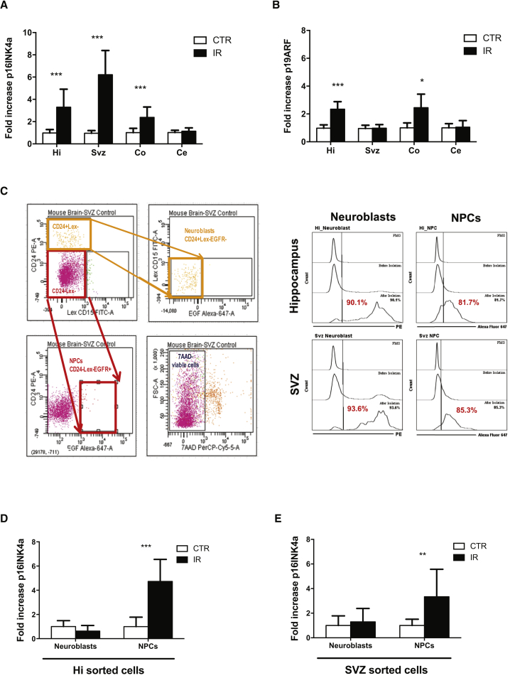
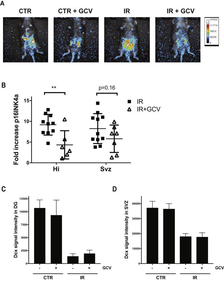
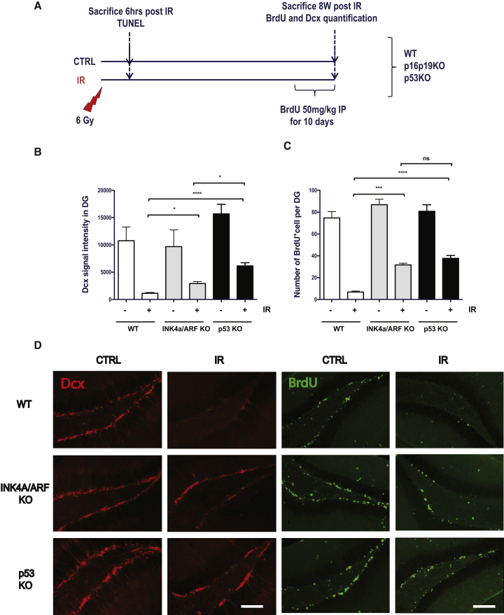
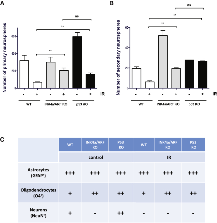
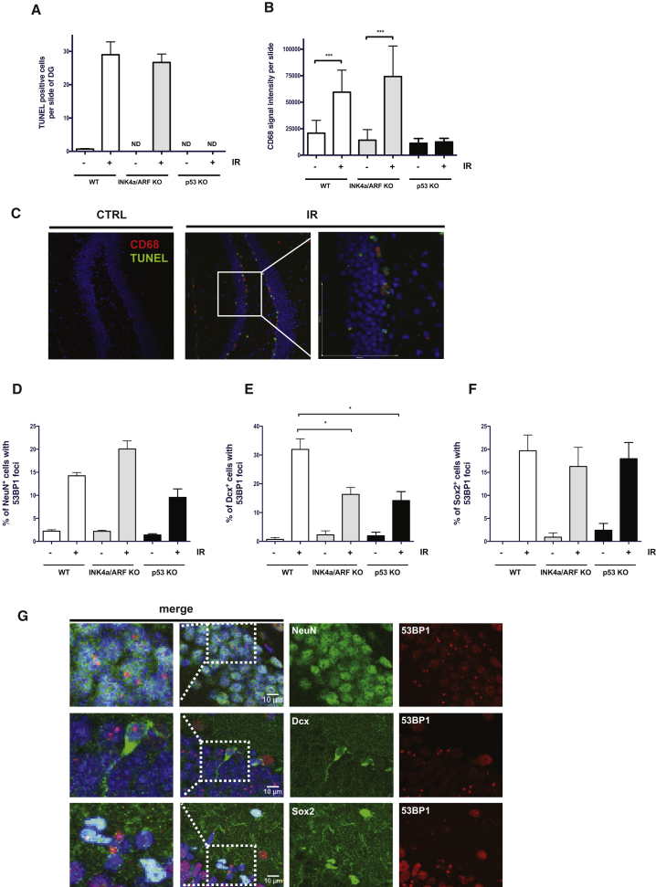
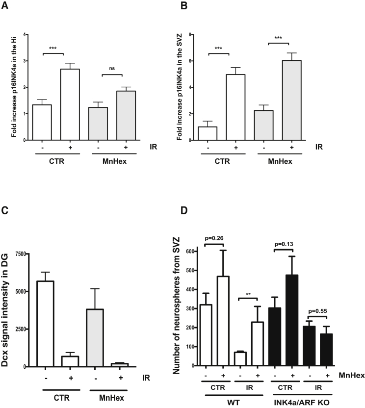
Brain neurogenesis is severely impaired following exposure to ionizing radiation (IR). We and others have shown that the expression of the tumor suppressor gene p16INK4a is increased in tissues exposed to IR and thus hypothesized that its expression could limit neurogenesis in the irradiated brain. Here, we found that exposure to IR leads to persistent DNA damage and the expression of p16INK4a in the hippocampus and subventricular zone regions. This was accompanied by a decline in neurogenesis, as determined by doublecortin expression and bromodeoxyuridine incorporation, an effect partially restored in Ink4a/arf-null mice. Increased neurogenesis in the absence of INK4a/ARF expression was independent of apoptosis and activation of the microglia. Moreover, treatment of irradiated mice with a superoxide dismutase mimetic or clearance of p16INK4a-expressing cells using mouse genetics failed to increase neurogenesis. In conclusion, our results suggest that IR-induced p16INK4a expression is a mechanism that limits neurogenesis.
脑神经发生在暴露于电离辐射(IR)后严重受损。我们和其他人已经表明,肿瘤抑制基因 p16INK4a 的表达在暴露于 IR 的组织中增加,因此我们假设其表达可能限制照射后脑中的神经发生。在这里,我们发现 IR 暴露导致 DNA 损伤和海马和脑室下区中 p16INK4a 的表达持续存在。这伴随着神经发生的下降,如双皮质素表达和溴脱氧尿苷掺入所确定的,在 Ink4a/arf 缺失小鼠中部分恢复。在没有 INK4a/ARF 表达的情况下增加的神经发生与细胞凋亡和小胶质细胞的激活无关。此外,用超氧化物歧化酶类似物处理照射的小鼠或使用小鼠遗传学清除表达 p16INK4a 的细胞未能增加神经发生。总之,我们的结果表明,IR 诱导的 p16INK4a 表达是限制神经发生的一种机制。