Institute of Clinical Physiology, Italian National Research Council, Pisa, Italy.
Institute of Life Sciences, Scuola Superiore Sant'Anna, Pisa, Italy.
J Diabetes Res. 2018 Mar 8;2018:4561309. doi: 10.1155/2018/4561309. eCollection 2018.
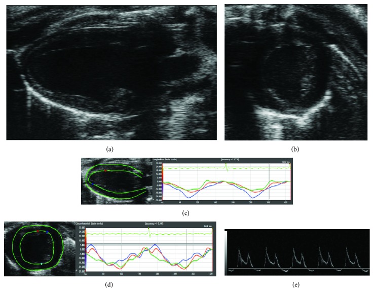
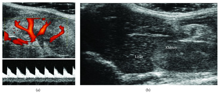
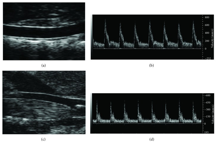
The availability of an animal model able to reliably mirror organ damage occurring in metabolic diseases is an urgent need. These models, mostly rodents, have not been fully characterized in terms of cardiovascular, renal, and hepatic ultrasound parameters, and only sparse values can be found in literature. Aim of this paper is to provide a detailed, noninvasive description of the heart, vessels, liver, and kidneys of the mouse by ultrasound imaging. Sixteen wild type and thirty-four male mice (11-week-old) were studied. State-of-the-art ultrasound technology was used to acquire images of cardiovascular, renal, and hepatic districts. A set of parameters describing function of the selected organs was evaluated. mice are characterized by systolic and diastolic dysfunction, confirmed by strain analysis. Abdominal aortic and carotid stiffness do not seem to be increased in diabetic rodents; furthermore, they are characterized by a smaller mean diameter for both vessels. Renal microcirculation is significantly compromised, while liver steatosis is only slightly higher in mice than in controls. We offer here for the first time an detailed ultrasonographic characterization of the mouse, providing a useful tool for a thoughtful choice of the right rodent model for any experimental design.
迫切需要有一种能够可靠反映代谢性疾病中器官损伤的动物模型。这些模型主要是啮齿动物,其心血管、肾脏和肝脏的超声参数尚未得到充分描述,文献中只有少量数据。本文旨在通过超声成像对小鼠的心脏、血管、肝脏和肾脏进行详细、无创的描述。研究了 16 只野生型和 34 只雄性小鼠(11 周龄)。使用最先进的超声技术获取心血管、肾脏和肝脏区域的图像。评估了一组描述所选器官功能的参数。糖尿病小鼠表现出收缩和舒张功能障碍,这通过应变分析得到了证实。腹主动脉和颈动脉的僵硬度似乎没有在糖尿病啮齿动物中增加;此外,它们的血管平均直径较小。肾脏微循环明显受损,而肝脏脂肪变性在 小鼠中比对照组仅略高。我们首次提供了对 小鼠的详细超声特征描述,为任何实验设计选择合适的啮齿动物模型提供了有用的工具。