• 文献检索
  • 文档翻译
  • 深度研究
  • 学术资讯
  • Suppr Zotero 插件Zotero 插件
  • 邀请有礼
  • 套餐&价格
  • 历史记录
应用&插件
Suppr Zotero 插件Zotero 插件浏览器插件Mac 客户端Windows 客户端微信小程序
定价
高级版会员购买积分包购买API积分包
服务
文献检索文档翻译深度研究API 文档MCP 服务
关于我们
关于 Suppr公司介绍联系我们用户协议隐私条款
关注我们

Suppr 超能文献

核心技术专利:CN118964589B侵权必究
粤ICP备2023148730 号-1Suppr @ 2026

文献检索

告别复杂PubMed语法,用中文像聊天一样搜索,搜遍4000万医学文献。AI智能推荐,让科研检索更轻松。

立即免费搜索

文件翻译

保留排版,准确专业,支持PDF/Word/PPT等文件格式,支持 12+语言互译。

免费翻译文档

深度研究

AI帮你快速写综述,25分钟生成高质量综述,智能提取关键信息,辅助科研写作。

立即免费体验

用于乳腺成像的多模态超声断层扫描:临床可行性的前瞻性研究

Multimodal ultrasound tomography for breast imaging: a prospective study of clinical feasibility.

作者信息

Forte S, Dellas S, Stieltjes B, Bongartz B

机构信息

University of Basel Hospital, Clinic for Radiology and Nuclear Medicine, Petersgraben 4, 4031 Basel, Switzerland.

出版信息

Eur Radiol Exp. 2017;1(1):27. doi: 10.1186/s41747-017-0029-y. Epub 2017 Dec 22.

DOI:10.1186/s41747-017-0029-y
PMID:29708167
原文链接:https://pmc.ncbi.nlm.nih.gov/articles/PMC5909358/
Abstract

BACKGROUND

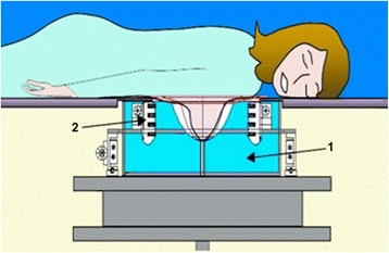
To describe the clinical set-up and evaluate the feasibility of multimodal ultrasound tomography (MUT) for breast imaging.

METHODS

Thirty-two consecutive patients referred for breast imaging and 24 healthy volunteers underwent MUT. In the 32 patients, the examination discomfort was compared to that of mammography (n = 31), handheld ultrasound (HUS) (n = 27) and magnetic resonance imaging (MRI) (n = 4) on a scale from 1 (lowest discomfort) to 10 (highest discomfort). MUT investigation time was recorded. Findings automatically detected by MUT were correlated with conventional imaging and biopsy results.

RESULTS

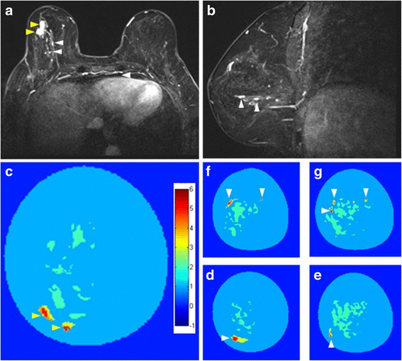
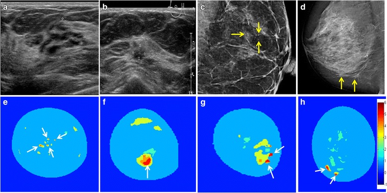
Breast MUT was well tolerated by all 56 participants; 55 bilateral exams were uneventful. During one exam, the digitalisation card failed and the exam was successfully repeated within three days. Mean examination discomfort was 1.6 (range = 1-5) for MUT, 1.5 (range = 1-5) for HUS, 5.3 (range = 3-7) for MRI, and 6.3 (range = 1-10) for mammography. MUT examination time was 38 ± 6 min (mean ± standard deviation). In the patients referred for breast imaging, MUT detected four lesions and indicated malignancy in three of these cases. These findings were confirmed by additional imaging and biopsy.

CONCLUSION

MUT is feasible in a clinical context considering examination time and patient acceptance. These interesting initial diagnostic findings warrant further studies.

摘要

背景

描述临床设置并评估多模态超声断层扫描(MUT)用于乳腺成像的可行性。

方法

32例连续转诊进行乳腺成像的患者和24名健康志愿者接受了MUT检查。在这32例患者中,将MUT检查的不适感与乳腺X线摄影(n = 31)、手持超声(HUS)(n = 27)和磁共振成像(MRI)(n = 4)的不适感进行比较,范围从1(最低不适感)到10(最高不适感)。记录MUT检查时间。MUT自动检测到的结果与传统成像和活检结果相关。

结果

所有56名参与者对乳腺MUT的耐受性良好;55例双侧检查均顺利。在一次检查中,数字化卡出现故障,该检查在三天内成功重复进行。MUT的平均检查不适感为1.6(范围 = 1 - 5),HUS为1.5(范围 = 1 - 5),MRI为5.3(范围 = 3 - 7),乳腺X线摄影为6.3(范围 = 1 - 10)。MUT检查时间为38±6分钟(平均值±标准差)。在转诊进行乳腺成像的患者中,MUT检测到4个病变,其中3例提示为恶性。这些结果通过额外的成像和活检得到证实。

结论

考虑到检查时间和患者接受度,MUT在临床环境中是可行的。这些有趣的初步诊断结果值得进一步研究。

https://cdn.ncbi.nlm.nih.gov/pmc/blobs/9d70/6091701/5880b9248a58/41747_2017_29_Fig3_HTML.jpg
https://cdn.ncbi.nlm.nih.gov/pmc/blobs/9d70/6091701/44a2dcd0488c/41747_2017_29_Fig1_HTML.jpg
https://cdn.ncbi.nlm.nih.gov/pmc/blobs/9d70/6091701/635e935e0d95/41747_2017_29_Fig2_HTML.jpg
https://cdn.ncbi.nlm.nih.gov/pmc/blobs/9d70/6091701/5880b9248a58/41747_2017_29_Fig3_HTML.jpg
https://cdn.ncbi.nlm.nih.gov/pmc/blobs/9d70/6091701/44a2dcd0488c/41747_2017_29_Fig1_HTML.jpg
https://cdn.ncbi.nlm.nih.gov/pmc/blobs/9d70/6091701/635e935e0d95/41747_2017_29_Fig2_HTML.jpg
https://cdn.ncbi.nlm.nih.gov/pmc/blobs/9d70/6091701/5880b9248a58/41747_2017_29_Fig3_HTML.jpg

相似文献

1
Multimodal ultrasound tomography for breast imaging: a prospective study of clinical feasibility.用于乳腺成像的多模态超声断层扫描:临床可行性的前瞻性研究
Eur Radiol Exp. 2017;1(1):27. doi: 10.1186/s41747-017-0029-y. Epub 2017 Dec 22.
2
Differentiation of BIRADS-4 small breast lesions via Multimodal Ultrasound Tomography.基于多模态超声层析成像技术对 BI-RADS-4 类小乳腺病灶的鉴别诊断
Eur Radiol. 2015 Feb;25(2):410-8. doi: 10.1007/s00330-014-3415-3. Epub 2014 Sep 14.
3
4
The role of magnetic resonance imaging in patients with palpable breast abnormalities and negative mammographic and sonographic findings.磁共振成像在可触及乳腺异常且乳腺 X 线摄影和超声检查结果阴性患者中的作用。
Breast J. 2020 Jul;26(7):1289-1295. doi: 10.1111/tbj.13793. Epub 2020 Feb 27.
5
Novel technology of multimodal ultrasound tomography detects breast lesions.多模态超声层析成像新技术检测乳腺病变。
Eur Radiol. 2013 Mar;23(3):673-83. doi: 10.1007/s00330-012-2659-z. Epub 2012 Sep 16.
6
Initial clinical results with a fusion prototype for mammography and three-dimensional ultrasound with a standard mammography system and a standard ultrasound probe.使用融合原型机结合标准乳腺摄影系统和标准超声探头进行乳腺摄影和三维超声检查的初步临床结果。
Acta Radiol. 2018 Dec;59(12):1406-1413. doi: 10.1177/0284185118762249. Epub 2018 Mar 2.
7
Surveillance of BRCA1 and BRCA2 mutation carriers with magnetic resonance imaging, ultrasound, mammography, and clinical breast examination.对携带BRCA1和BRCA2基因突变者进行磁共振成像、超声、乳房X线摄影及临床乳腺检查监测。
JAMA. 2004 Sep 15;292(11):1317-25. doi: 10.1001/jama.292.11.1317.
8
Contrast-enhanced magnetic resonance mammography: does it affect surgical decision-making in patients with breast cancer?对比增强磁共振乳腺成像:它会影响乳腺癌患者的手术决策吗?
Breast Cancer Res Treat. 2007 Nov;106(1):65-74. doi: 10.1007/s10549-006-9472-9. Epub 2007 Jan 3.
9
The challenge of imaging dense breast parenchyma: is magnetic resonance mammography the technique of choice? A comparative study with x-ray mammography and whole-breast ultrasound.致密乳腺实质成像的挑战:磁共振乳腺造影是首选技术吗?一项与乳腺X线摄影和全乳超声的对比研究。
Invest Radiol. 2009 Jul;44(7):412-21. doi: 10.1097/RLI.0b013e3181a53654.
10
Initial results of the FUSION-X-US prototype combining 3D automated breast ultrasound and digital breast tomosynthesis.融合 X-US 原型机的初步结果,该原型机结合了三维自动化乳腺超声和数字乳腺断层合成技术。
Eur Radiol. 2018 Jun;28(6):2499-2506. doi: 10.1007/s00330-017-5235-8. Epub 2018 Jan 4.

引用本文的文献

1
Dynamic Speed of Sound Adaptive Transmission-Reflection Ultrasound Computed Tomography.动态声速自适应透射-反射超声计算机断层成像技术。
Sensors (Basel). 2023 Apr 3;23(7):3701. doi: 10.3390/s23073701.
2
Ultrasound and Photoacoustic Imaging of Breast Cancer: Clinical Systems, Challenges, and Future Outlook.乳腺癌的超声与光声成像:临床系统、挑战及未来展望
J Clin Med. 2022 Feb 22;11(5):1165. doi: 10.3390/jcm11051165.
3
Transmission-reflection optoacoustic ultrasound (TROPUS) computed tomography of small animals.小动物的透射-反射光声超声(TROPUS)计算机断层扫描

本文引用的文献

1
Review of radiation dose estimates in digital breast tomosynthesis relative to those in two-view full-field digital mammography.数字化乳腺断层合成与双视图全视野数字化乳腺摄影的辐射剂量估计值综述。
Breast. 2015 Apr;24(2):93-9. doi: 10.1016/j.breast.2014.12.002. Epub 2014 Dec 29.
2
Differentiation of BIRADS-4 small breast lesions via Multimodal Ultrasound Tomography.基于多模态超声层析成像技术对 BI-RADS-4 类小乳腺病灶的鉴别诊断
Eur Radiol. 2015 Feb;25(2):410-8. doi: 10.1007/s00330-014-3415-3. Epub 2014 Sep 14.
3
Novel technology of multimodal ultrasound tomography detects breast lesions.
Light Sci Appl. 2019 Jan 30;8:18. doi: 10.1038/s41377-019-0130-5. eCollection 2019.
多模态超声层析成像新技术检测乳腺病变。
Eur Radiol. 2013 Mar;23(3):673-83. doi: 10.1007/s00330-012-2659-z. Epub 2012 Sep 16.
4
Radiologists' performance in the detection of benign and malignant masses with 3D automated breast ultrasound (ABUS).放射科医生使用 3D 自动乳腺超声 (ABUS) 检测良性和恶性肿块的表现。
Eur J Radiol. 2011 Apr;78(1):99-103. doi: 10.1016/j.ejrad.2011.01.074. Epub 2011 Feb 16.
5
Breast cancer detection: radiologists' performance using mammography with and without automated whole-breast ultrasound.乳腺癌检测:使用配有和不配有自动全乳房超声的乳腺 X 线摄影术时放射科医生的表现。
Eur Radiol. 2010 Nov;20(11):2557-64. doi: 10.1007/s00330-010-1844-1. Epub 2010 Jul 15.
6
Breast cancer detection using automated whole breast ultrasound and mammography in radiographically dense breasts.乳腺 X 线摄影致密型乳腺中应用自动全乳腺超声和乳腺 X 线摄影筛查乳腺癌。
Eur Radiol. 2010 Mar;20(3):734-42. doi: 10.1007/s00330-009-1588-y. Epub 2009 Sep 2.
7
Combined screening with ultrasound and mammography vs mammography alone in women at elevated risk of breast cancer.超声与乳腺X线摄影联合筛查与单纯乳腺X线摄影筛查对乳腺癌高危女性的效果比较
JAMA. 2008 May 14;299(18):2151-63. doi: 10.1001/jama.299.18.2151.
8
Differentiation of cancerous lesions in excised human breast specimens using multiband attenuation profiles from ultrasonic transmission tomography.利用超声透射断层扫描的多波段衰减剖面鉴别切除的人体乳腺标本中的癌性病变。
J Ultrasound Med. 2008 Mar;27(3):435-51. doi: 10.7863/jum.2008.27.3.435.
9
Segmentation methodology for automated classification and differentiation of soft tissues in multiband images of high-resolution ultrasonic transmission tomography.用于高分辨率超声透射断层扫描多波段图像中软组织自动分类与区分的分割方法
IEEE Trans Med Imaging. 2006 Aug;25(8):1068-78. doi: 10.1109/tmi.2006.877443.
10
Effect of screening and adjuvant therapy on mortality from breast cancer.筛查与辅助治疗对乳腺癌死亡率的影响。
N Engl J Med. 2005 Oct 27;353(17):1784-92. doi: 10.1056/NEJMoa050518.