Kratkiewicz Karl, Pattyn Alexander, Alijabbari Naser, Mehrmohammadi Mohammad
Department of Oncology, Wayne State University, Detroit, MI 48202, USA.
Department of Biomedical Engineering, Wayne State University, Detroit, MI 48202, USA.
J Clin Med. 2022 Feb 22;11(5):1165. doi: 10.3390/jcm11051165.
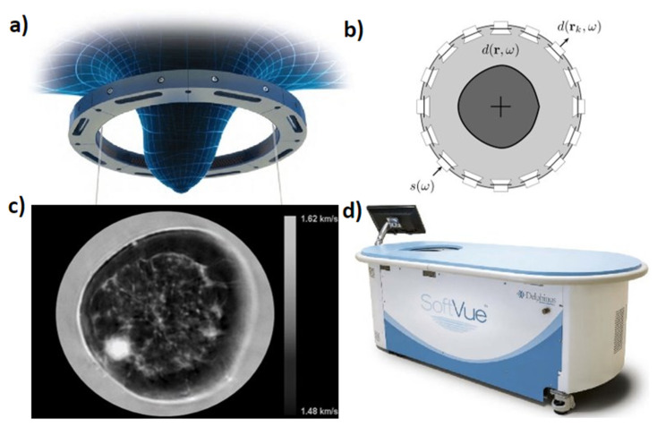
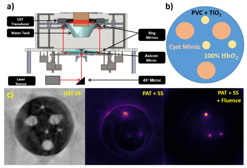
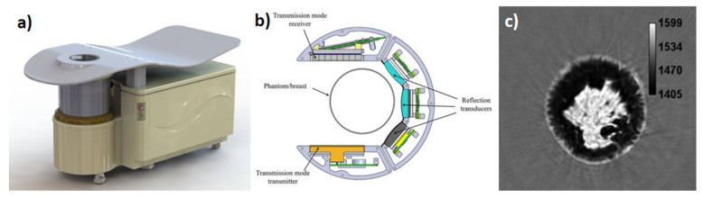
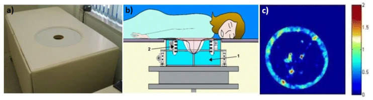
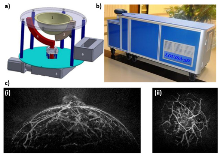
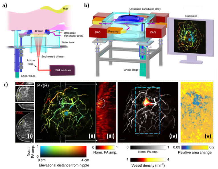
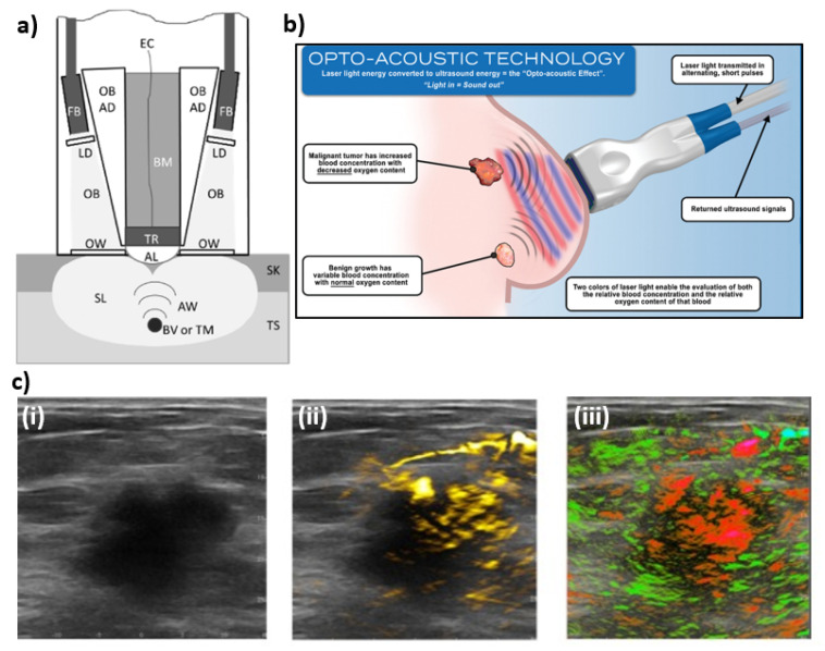
Presently, breast cancer diagnostic methods are dominated by mammography. Although drawbacks of mammography are present including ionizing radiation and patient discomfort, not many alternatives are available. Ultrasound (US) is another method used in the diagnosis of breast cancer, commonly performed on women with dense breasts or in differentiating cysts from solid tumors. Handheld ultrasound (HHUS) and automated breast ultrasound (ABUS) are presently used to generate reflection images which do not contain quantitative information about the tissue. This limitation leads to a subjective interpretation from the sonographer. To rectify the subjective nature of ultrasound, ultrasound tomography (UST) systems have been developed to acquire both reflection and transmission UST (TUST) images. This allows for quantitative assessment of tissue sound speed (SS) and acoustic attenuation which can be used to evaluate the stiffness of the lesions. Another imaging modality being used to detect breast cancer is photoacoustic tomography (PAT). Utilizing much of the same hardware as ultrasound tomography, PAT receives acoustic waves generated from tissue chromophores that are optically excited by a high energy pulsed laser. This allows the user to ideally produce chromophore concentration maps or extract other tissue parameters through spectroscopic PAT. Here, several systems in the area of TUST and PAT are discussed along with their advantages and disadvantages in breast cancer diagnosis. This overview of available systems can provide a landscape of possible intersections and future refinements in cancer diagnosis.
目前,乳腺癌诊断方法以乳房X线摄影为主。尽管乳房X线摄影存在一些缺点,包括电离辐射和患者不适,但可用的替代方法并不多。超声(US)是用于乳腺癌诊断的另一种方法,常用于乳房致密的女性或用于区分囊肿与实体瘤。目前,手持式超声(HHUS)和自动乳腺超声(ABUS)用于生成不包含组织定量信息的反射图像。这种局限性导致超声检查人员的主观解读。为了纠正超声的主观性,已经开发了超声断层扫描(UST)系统来获取反射和透射UST(TUST)图像。这允许对组织声速(SS)和声学衰减进行定量评估,可用于评估病变的硬度。另一种用于检测乳腺癌的成像模态是光声断层扫描(PAT)。PAT使用与超声断层扫描大部分相同的硬件,接收由组织发色团产生的声波,这些发色团由高能脉冲激光进行光学激发。这允许用户理想地生成发色团浓度图或通过光谱PAT提取其他组织参数。在此,讨论了TUST和PAT领域的几种系统及其在乳腺癌诊断中的优缺点。对现有系统的这一概述可以提供癌症诊断中可能的交叉点和未来改进的概况。