Department of Obstetrics & Gynecology, Southwest Hospital, Third Military Medical University, Chongqing, 400038, China.
Bjrigham Young University, ID 272 Rigby Hall, Rexburg, 83460-4500, USA.
J Ovarian Res. 2018 May 2;11(1):36. doi: 10.1186/s13048-018-0403-2.
Ovarian cancer stem cells (OCSCs) contribute to the poor prognosis of ovarian cancer. Involvement of the androgen receptor (AR) in the malignant behaviors of other tumors has been reported. However, whether AR associates with Nanog (a stem cell marker) and participates in OCSC functions remain unclear. In this study, we investigated the interaction of Nanog with AR and examined whether this interaction induced stem-like properties in ovarian cancer cells.
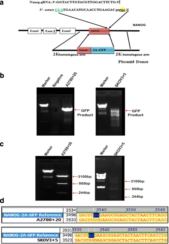
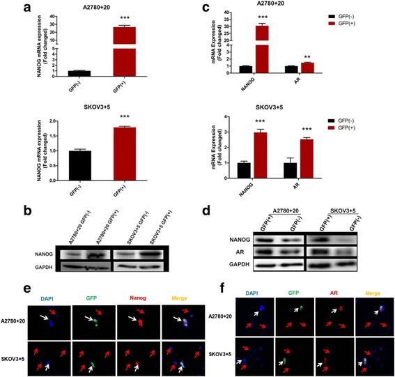
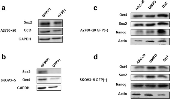
AR and Nanog expression in ovarian tumors was evaluated. Using the CRISPR/Cas9 system, we constructed a Nanog green fluorescent protein (GFP) marker cell model to investigate the expression and co-localization of Nanog and AR. Then, we examined the effect of androgen on the Nanog promoter in ovarian cancer cell lines (A2780 and SKOV3). After androgen or anti-androgen treatment, cell proliferation, migration, sphere formation, colony formation and tumorigenesis were assessed in vitro and in vivo.
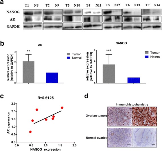
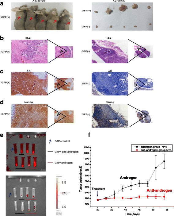
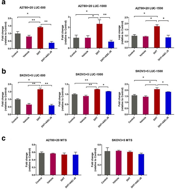
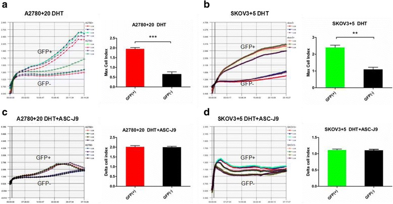
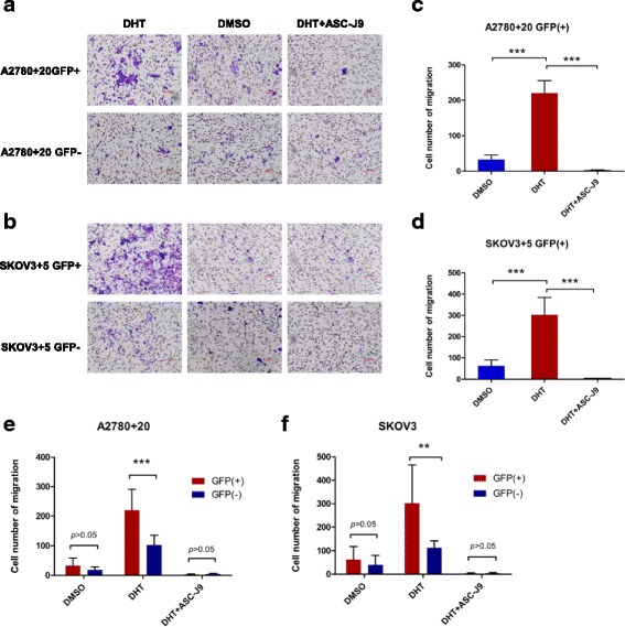
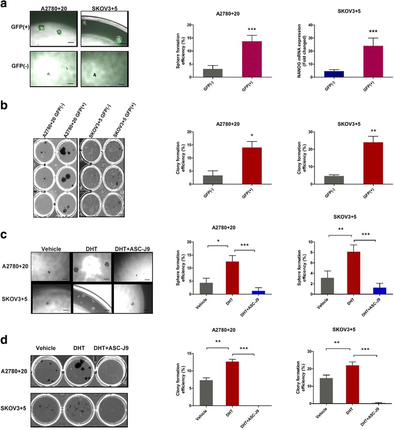
Both AR and Nanog expression were obviously high in ovarian tumors. Our results showed that Nanog expression was correlated with AR expression. The androgen 5α-dihydrotestosterone (DHT) activated Nanog promoter transcription. Meanwhile, Nanog GFP-positive cells treated with DHT exhibited higher levels of proliferation, migration, sphere formation and colony formation. We also observed that the tumorigenesis of Nanog GFP-positive cells was significantly higher than that of the GFP-negative cells. Xenografts of Nanog GFP-positive cells showed significant differences when treated with androgen or anti-androgen drugs in vivo.
The interaction of Nanog with the AR signaling axis might induce or contribute to OCSC regulation. In addition, androgen might promote stemness characteristics in ovarian cancer cells by activating the Nanog promoter. This finding merits further study because it may provide a new understanding of OCSC regulation from a hormone perspective and lead to the reevaluation of stem cell therapy for ovarian cancer.
卵巢癌干细胞(OCSCs)导致卵巢癌预后不良。雄激素受体(AR)参与其他肿瘤的恶性行为已有报道。然而,AR 是否与 Nanog(干细胞标志物)相互作用并参与 OCSC 功能尚不清楚。在这项研究中,我们研究了 Nanog 与 AR 的相互作用,并研究了这种相互作用是否诱导卵巢癌细胞产生干细胞样特性。
评估卵巢肿瘤中 AR 和 Nanog 的表达。我们使用 CRISPR/Cas9 系统构建了 Nanog 绿色荧光蛋白(GFP)标记细胞模型,以研究 Nanog 和 AR 的表达和共定位。然后,我们研究了雄激素对卵巢癌细胞系(A2780 和 SKOV3)中 Nanog 启动子的影响。在雄激素或抗雄激素治疗后,在体外和体内评估细胞增殖、迁移、球体形成、集落形成和肿瘤发生。
卵巢肿瘤中 AR 和 Nanog 的表达均明显升高。我们的结果表明,Nanog 的表达与 AR 的表达相关。雄激素 5α-二氢睾酮(DHT)激活了 Nanog 启动子转录。同时,用 DHT 处理的 Nanog GFP 阳性细胞表现出更高水平的增殖、迁移、球体形成和集落形成。我们还观察到,Nanog GFP 阳性细胞的肿瘤发生明显高于 GFP 阴性细胞。体内,用雄激素或抗雄激素药物处理 Nanog GFP 阳性细胞的异种移植物时,差异显著。
Nanog 与 AR 信号轴的相互作用可能诱导或有助于 OCSC 调节。此外,雄激素可能通过激活 Nanog 启动子促进卵巢癌细胞的干性特征。这一发现值得进一步研究,因为它可能从激素角度提供对 OCSC 调节的新认识,并导致对卵巢癌干细胞治疗的重新评估。