Menceva Zaklina, Dimitrovski Oliver, Popovska Mirjana, Spasovski Spiro, Spirov Vancho, Petrushevska Gordana
Department of Oral Surgery, University Dental Clinical Centre St. Pantelejmon, Faculty of Dentistry, Ss Cyril and Methodius University of Skopje, Skopje, Republic of Macedonia.
Department of Oral Surgery and Implantology, Faculty of Dentistry, Ss. Cyril and Methodius University of Skopje, Skopje, Republic of Macedonia.
Open Access Maced J Med Sci. 2018 Mar 27;6(4):675-679. doi: 10.3889/oamjms.2018.127. eCollection 2018 Apr 15.
The correction of the gingival recession is of esthetical and functional significance, but the tissue regeneration can only be confirmed by a histological examination.
This study aims to make a comparison between the free gingival graft and the autograft.
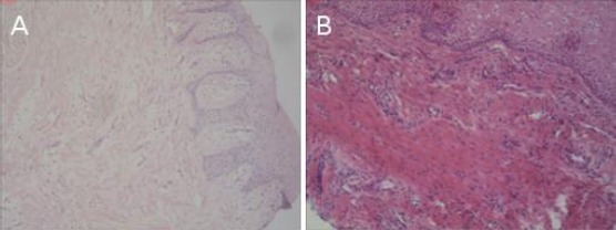
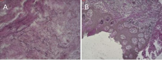
This study included 24 patients with single and multiple gingival recessions. Twelve patients were treated with a free gingival graft and the other twelve with a micrograft. Six months after the surgical procedure, a micro-punch biopsy of the transplantation area was performed. The tissue was histologically evaluated, graded in 4 categories: immature, mature, fragmented and edematous collagen tissue. The elastic fibres were also examined and graded in three categories: with a normal structure, fragmented rare and fragmented multiplied.
Regarding the type of collagen tissue that was present, there was a significant difference between the two groups of patients, with a larger number of patients treated with a micrograft showing a presence of mature tissue, compared to the patients treated with a free gingival graft. A larger number of patients in both of the groups displayed elastic fibres with a rare fragmented structure; 33.3% of the patients showed a normal structure; 50% demonstrated a normal structure.
The patients treated with a free gingival graft showed a larger presence of fragmented collagen tissue and fragmented elastic fibres, whereas a mature tissue was predominantly present in the surgical area where a Geistlich Mucograft was placed.
牙龈退缩的矫正具有美学和功能意义,但组织再生只能通过组织学检查来确认。
本研究旨在比较游离龈瓣移植术和自体移植术。
本研究纳入24例单发性和多发性牙龈退缩患者。12例患者接受游离龈瓣移植术治疗,另外12例接受微小移植术治疗。手术6个月后,对移植区域进行微型穿刺活检。对组织进行组织学评估,分为4类:未成熟、成熟、破碎和水肿的胶原组织。还对弹性纤维进行检查并分为3类:结构正常、罕见破碎和大量破碎。
关于存在的胶原组织类型,两组患者之间存在显著差异,与接受游离龈瓣移植术治疗的患者相比,接受微小移植术治疗的患者中显示成熟组织的人数更多。两组中均有较多患者的弹性纤维呈现罕见的破碎结构;33.3%的患者显示结构正常;50%的患者显示结构正常。
接受游离龈瓣移植术治疗的患者中破碎的胶原组织和破碎的弹性纤维较多,而放置Geistlich生物牙龈移植材料的手术区域主要存在成熟组织。