Hamidabadi Hatef Ghasemi, Shafaroudi Majid Malekzadeh, Seifi Morteza, Bojnordi Maryam Nazm, Behruzi Masume, Gholipourmalekabadi Mazaher, Shafaroudi Ali Malekzadeh, Rezaei Nourollah
Immunogenetic Research Center (IRC),Faculty of Medicine, Mazandaran University of Medical Sciences, Sari, Iran.
Department of Anatomy and Cell Biology, Faculty of Medicine, Mazandaran University of Medical Sciences, Sari, Iran.
Med Arch. 2018 Apr;72(2):88-93. doi: 10.5455/medarh.2018.72.88-93.
The repair of critical-sized defects (CSDs) are one of the most challenging orthopedic problems and the attempts for development of an ideal scaffold for treatment of large bone defect are ongoing.
The aim of this study was the effectiveness of hydroxyapatite-gelatin seeded with bone marrow stromal cells construct for healing of critical-sized bone defect in vivo.
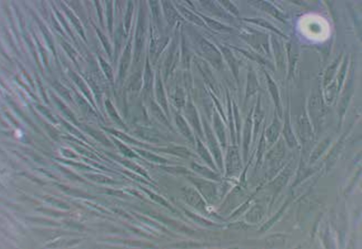
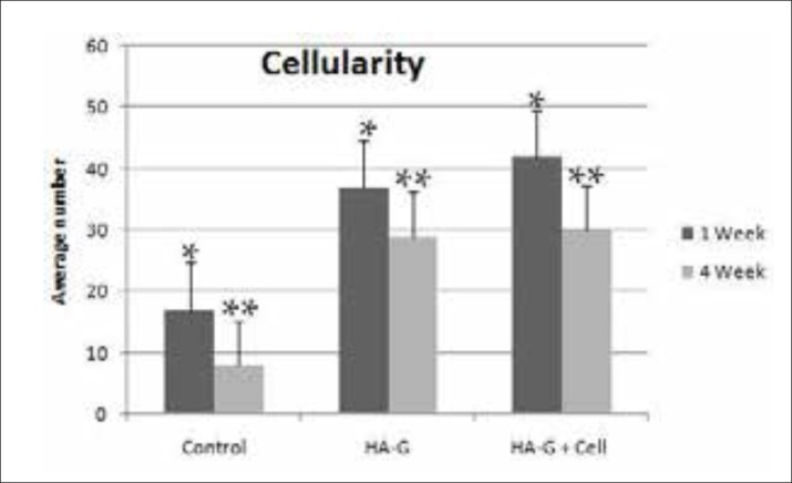
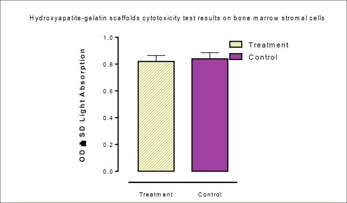
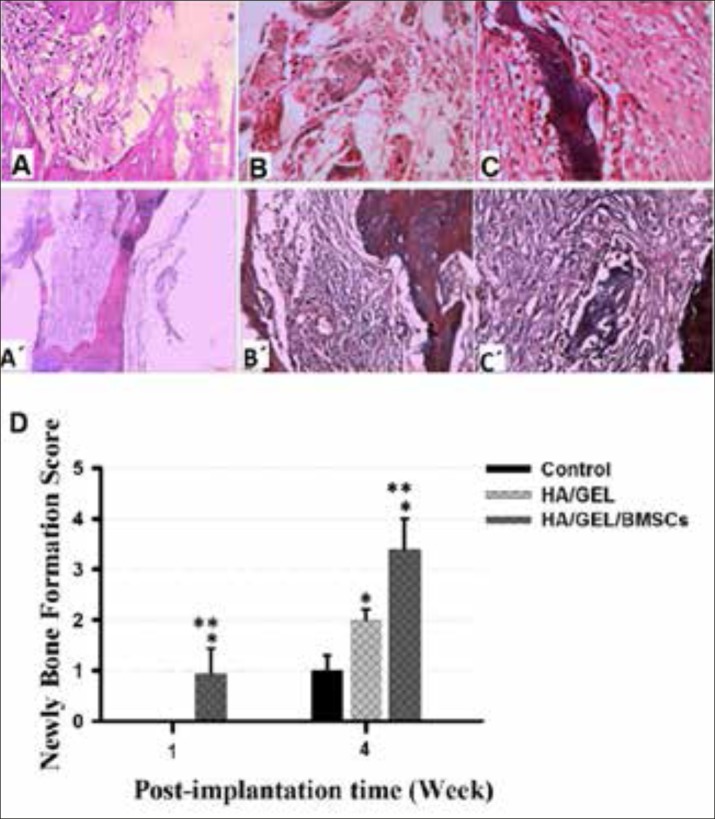
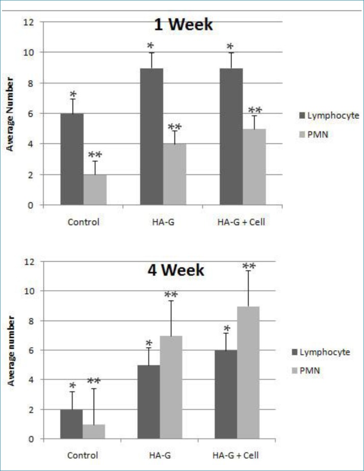
In this experimental study, the bone marrow stromal cells (BMSCs) were isolated by flushing method. For in vitro study, the cells were seeded on the scaffold and the cell viability as well as cytotoxicity were tested by MTT and LDH specific activity. The scaffold-cell construct was implanted into the critical-sized bone defect created in calvaria of Wistar male rats.15 rats were randomly divided into 3 groups (n=5), group 1 (control group): Injury without transplantation, group 2: implanted with hydroxyapatite-gelatin scaffold, group 3: hydroxyapatite-gelatin scaffold seeded with BMSCs. At different days post-implantation, the implanted site was collected and the bone healing was evaluated through H&E and Masson's Trichrome staining. ANOVA and paired t-test were used for data comparison and P<0.05 was considered significant.
The results of MTT showed that the scaffold has no toxic effects on stromal cells. The first signs of ossification in hydroxyapatite-gelatin with BMSCs cells group appeared in the first week. However, in the fourth week, ossification was completed and the scaffold remaining was found as embedded islands in the spongy bone tissue. The greatest number of lymphocytes in the experimental group was observed after one week of planting scaffold.
Hydroxyapatite-gelatin scaffold coated with BMSCs cells has a potential role in the healing process of bone and would be a possible new therapeutic strategy to repair extensive bone lesions.
临界尺寸骨缺损(CSDs)的修复是骨科领域最具挑战性的问题之一,目前仍在不断尝试开发用于治疗大骨缺损的理想支架。
本研究旨在探讨接种骨髓基质细胞的羟基磷灰石-明胶构建体在体内修复临界尺寸骨缺损的有效性。
在本实验研究中,采用冲洗法分离骨髓基质细胞(BMSCs)。在体外研究中,将细胞接种到支架上,并通过MTT和LDH比活性检测细胞活力和细胞毒性。将支架-细胞构建体植入Wistar雄性大鼠颅骨的临界尺寸骨缺损处。15只大鼠随机分为3组(n = 5),第1组(对照组):损伤后未进行移植;第2组:植入羟基磷灰石-明胶支架;第3组:接种BMSCs的羟基磷灰石-明胶支架。在植入后的不同天数,收集植入部位,通过苏木精-伊红(H&E)染色和马松三色染色评估骨愈合情况。采用方差分析和配对t检验进行数据比较,P<0.05认为具有统计学意义。
MTT结果显示,该支架对基质细胞无毒性作用。接种BMSCs细胞的羟基磷灰石-明胶组在第一周出现了骨化的最初迹象。然而,在第四周时,骨化完成,剩余的支架被发现为海绵骨组织中嵌入的岛状物。在植入支架一周后,实验组观察到的淋巴细胞数量最多。
接种BMSCs细胞的羟基磷灰石-明胶支架在骨愈合过程中具有潜在作用,可能是修复大面积骨损伤的一种新的治疗策略。