Santos German Iris Jasmin, Pomini Karina Torres, Bighetti Ana Carolina Cestari, Andreo Jesus Carlos, Reis Carlos Henrique Bertoni, Shinohara André Luis, Rosa Júnior Geraldo Marco, Teixeira Daniel de Bortoli, Rosso Marcelie Priscila de Oliveira, Buchaim Daniela Vieira, Buchaim Rogério Leone
Department of Biological Sciences (Anatomy), Bauru School of Dentistry, University of São Paulo (USP), Bauru, São Paulo 17012-901, Brazil.
Department of Dentistry, Faculty of Health Science, Universidad Iberoamericana (UNIBE), Santo Domingo 10203, Dominican Republic.
Materials (Basel). 2020 Feb 4;13(3):695. doi: 10.3390/ma13030695.
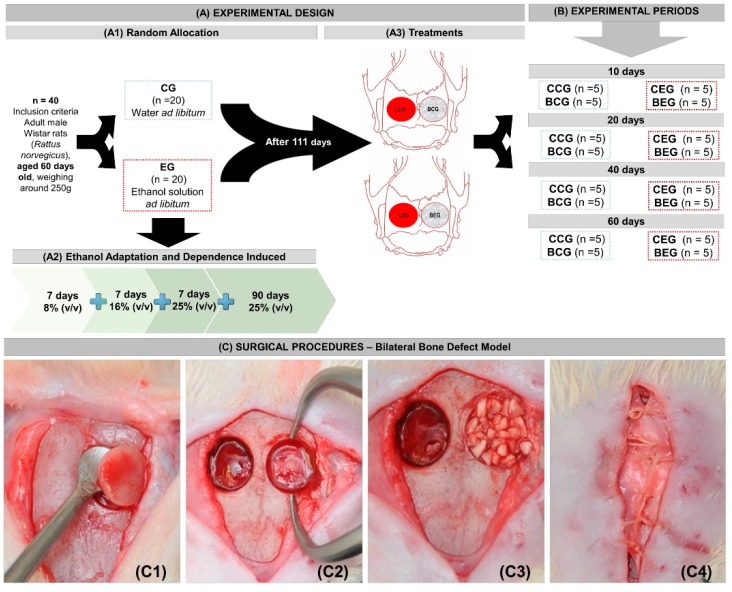
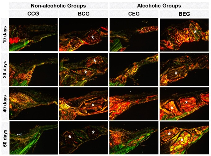
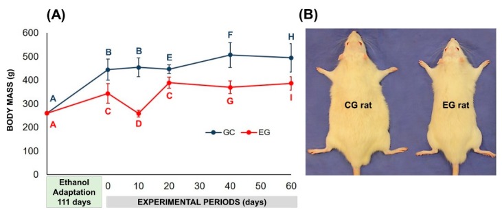
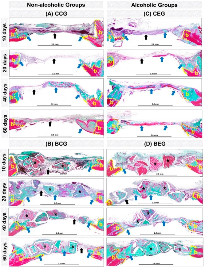
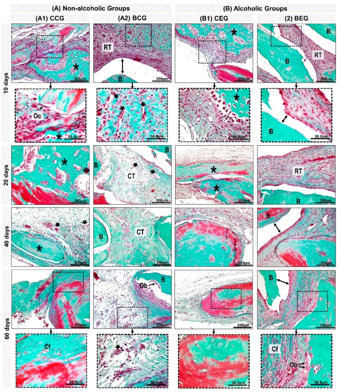
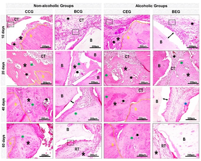
To assess the effects of chronic alcoholism on the repair of bone defects associated with xenograft. Forty male rats were distributed in: control group (CG, = 20) and experimental group (EG, = 20), which received 25% ethanol ad libitum after a period of adaptation. After 90 days of liquid diet, the rats were submitted to 5.0-mm bilateral craniotomy on the parietal bones, subdividing into groups: CCG (control group that received only water with liquid diet and the defect was filled with blood clot), BCG (control group that received only water with liquid diet and the defect was filled with biomaterial), CEG (alcoholic group that received only ethanol solution 25% / with liquid diet and the defect was filled with blood clot), and BEG (alcoholic group that received only ethanol solution 25% / with liquid diet and the defect was filled with biomaterial). In the analysis of body mass, the drunk animals presented the lowest averages in relation to non-drunk animals during the experimental period. Histomorphologically all groups presented bone formation restricted to the defect margins at 60 days, with bone islets adjacent to the BCG biomaterial particles. CEG showed significant difference compared to BEG only at 40 days (17.42 ± 2.78 vs. 9.59 ± 4.59, respectively). In the birefringence analysis, in early periods all groups showed red-orange birefringence turning greenish-yellow at the end of the experiment. The results provided that, regardless of clinical condition, i.e., alcoholic or non-alcoholic, in the final period of the experiment, the process of bone defect recomposition was similar with the use of xenograft or only clot.
评估慢性酒精中毒对与异种移植相关的骨缺损修复的影响。将40只雄性大鼠分为:对照组(CG,n = 20)和实验组(EG,n = 20),实验组在适应期后随意饮用25%乙醇。液体饮食90天后,对大鼠进行双侧顶骨5.0毫米开颅手术,再细分为:CCG(仅饮用含液体饮食的水且缺损用血凝块填充的对照组)、BCG(仅饮用含液体饮食的水且缺损用生物材料填充的对照组)、CEG(饮用25%乙醇溶液/含液体饮食且缺损用血凝块填充的酒精组)和BEG(饮用25%乙醇溶液/含液体饮食且缺损用生物材料填充的酒精组)。在体重分析中,在实验期间,醉酒动物的平均体重相对于未醉酒动物最低。组织形态学上,所有组在60天时骨形成均局限于缺损边缘,BCG组的生物材料颗粒附近有骨岛。CEG组与BEG组仅在40天时存在显著差异(分别为17.42±2.78和9.59±4.59)。在双折射分析中,早期所有组均显示红橙色双折射,在实验结束时变为绿黄色。结果表明,无论临床状况如何,即酒精性或非酒精性,在实验末期,使用异种移植或仅使用血凝块时骨缺损重组过程相似。