Mazzei Maria Antonietta, Bagnacci Giulio, Gentili Francesco, Nigri Andrea, Pelini Veronica, Vindigni Carla, Mazzei Francesco Giuseppe, Baiocchi Gian Luca, Pittiani Frida, Morgagni Paolo, Petrella Enrico, Mura Gianni, Verdelli Beatrice, Bencivenga Maria, Giacopuzzi Simone, Marrelli Daniele, Roviello Franco, Volterrani Luca
Department of Medical, Surgical and Neuro Sciences, Unit of Diagnostic Imaging, Azienda Ospedaliera Universitaria Senese, University of Siena, Siena, Italy.
Faculty of Statistics, Sapienza University of Rome, Roma, Italy.
Gastroenterol Res Pract. 2018 Mar 15;2018:1794524. doi: 10.1155/2018/1794524. eCollection 2018.
To investigate the role of maximum tumour diameter (D-max) reduction rate at CT examination in predicting histopathological tumour regression grade (TRG according to the Becker grade), after neoadjuvant chemotherapy (NAC), in patients with resectable advanced gastric cancer (AGC).
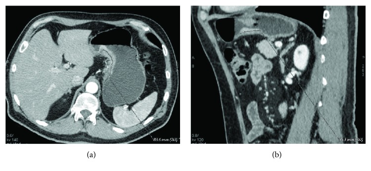
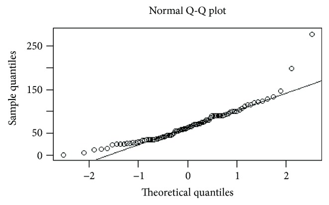
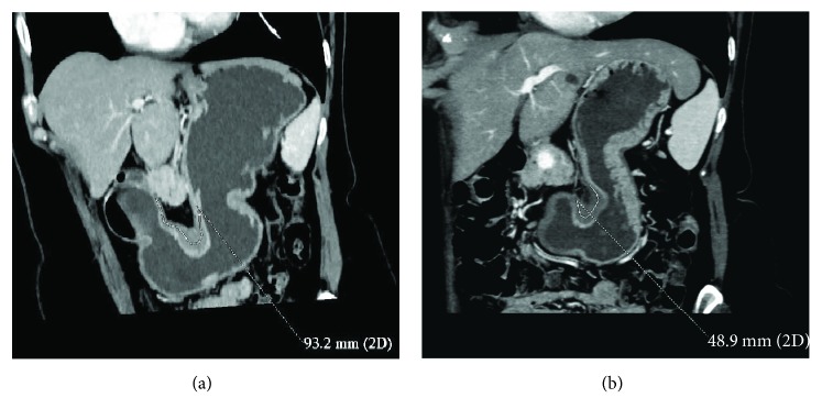
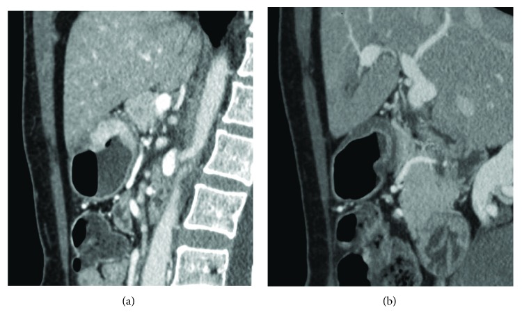
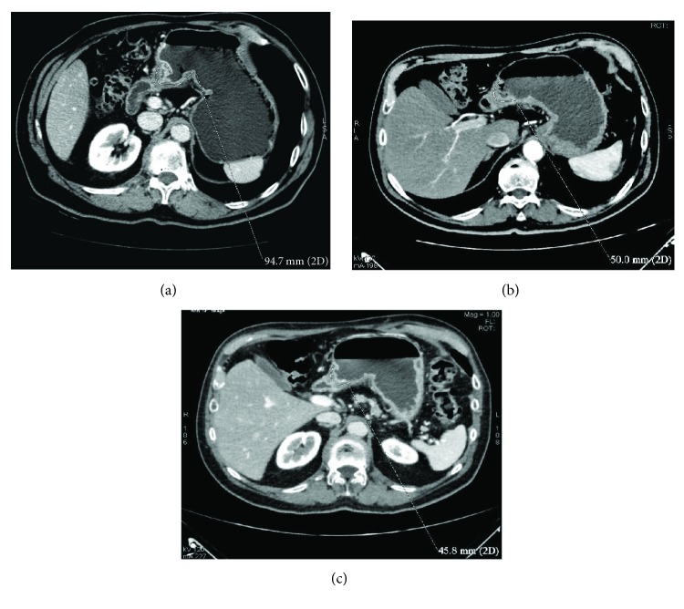
Eighty-six patients (53 M, mean age 62.1 years) with resectable AGC (≥T3 or N+), treated with NAC and radical surgery, were enrolled from 5 centres of the Italian Research Group for Gastric Cancer (GIRCG). Staging and restaging CT and histological results were retrospectively reviewed. CT examinations were contrast enhanced, and the stomach was previously distended. The D-max was measured using 2D software and compared with Becker TRG. Statistical data were obtained using "R" software.
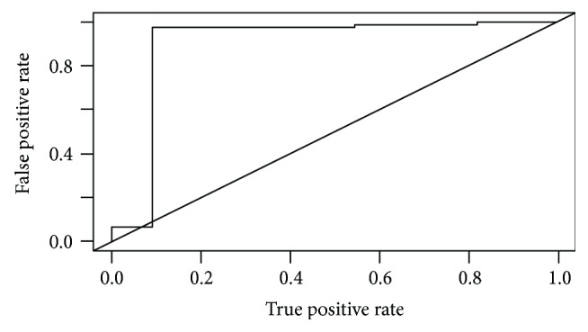
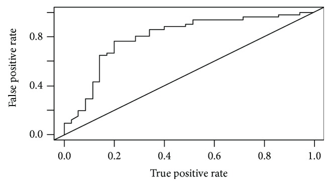
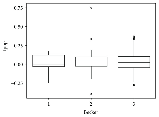
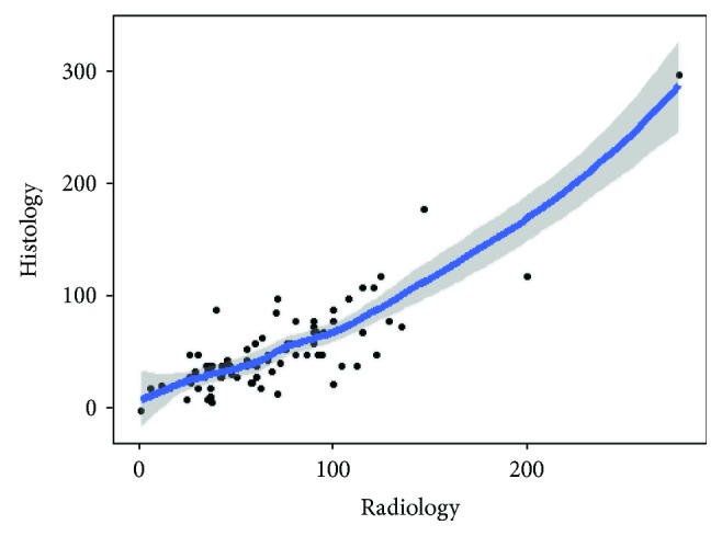
The interobserver agreement was good/very good. Becker TRG was predicted by CT with a sensitivity and specificity, respectively, of 97.3% and 90.9% for Becker 1 (D-max reduction rate > 65.1%), 76.4% and 80% for Becker 3 (D-max reduction rate < 29.9%), and 70.8% and 83.9% for Becker 2. Correlation between radiological and histological D-max measurements was strongly confirmed by the correlation index (c.i.= 0.829).
D-max reduction rate in AGC patients may be helpful as a simple and reproducible radiological index in predicting TRG after NAC.
探讨在可切除的进展期胃癌(AGC)患者中,新辅助化疗(NAC)后CT检查时肿瘤最大直径(D-max)缩小率在预测组织病理学肿瘤退缩分级(根据Becker分级的TRG)中的作用。
从意大利胃癌研究组(GIRCG)的5个中心纳入86例(53例男性,平均年龄62.1岁)可切除的AGC(≥T3或N+)患者,这些患者接受了NAC和根治性手术。对分期和再分期CT及组织学结果进行回顾性分析。CT检查采用增强扫描,检查前胃已充盈。使用二维软件测量D-max,并与Becker TRG进行比较。使用“R”软件获取统计数据。
观察者间一致性良好/非常好。CT对Becker TRG的预测,Becker 1级(D-max缩小率>65.1%)的敏感性和特异性分别为97.3%和90.9%,Becker 3级(D-max缩小率<29.9%)为76.4%和80%,Becker 2级为70.8%和83.9%。相关指数(c.i.=0.829)有力地证实了放射学和组织学D-max测量值之间的相关性。
AGC患者的D-max缩小率作为一种简单且可重复的放射学指标,可能有助于预测NAC后的TRG。