Faculty of Medicine, University of Southampton, Southampton, UK.
Biogen, Cambridge, USA.
Acta Neuropathol. 2018 Jul;136(1):139-152. doi: 10.1007/s00401-018-1862-7. Epub 2018 May 12.
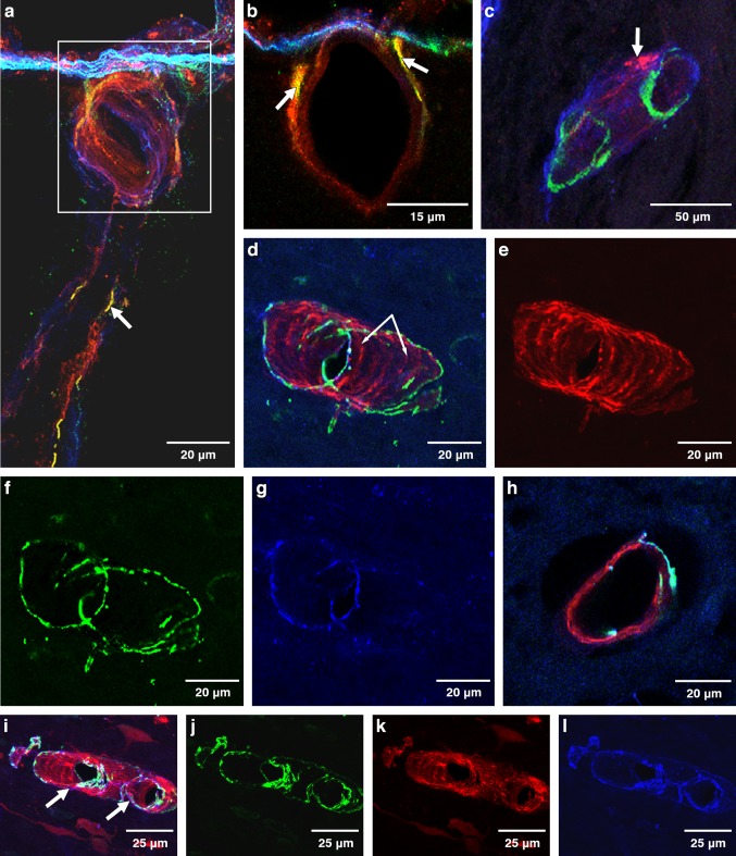
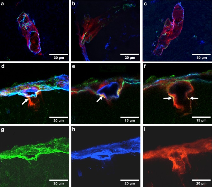
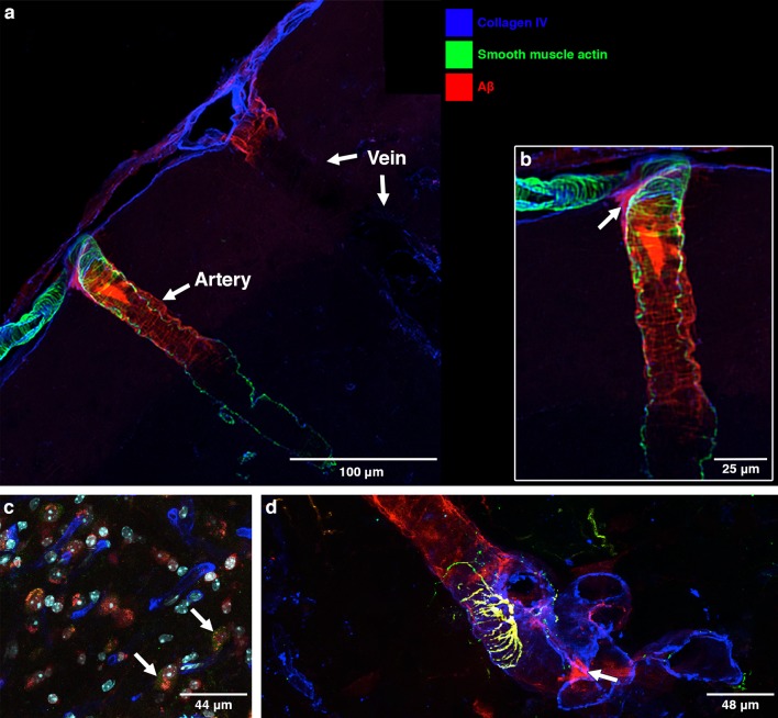
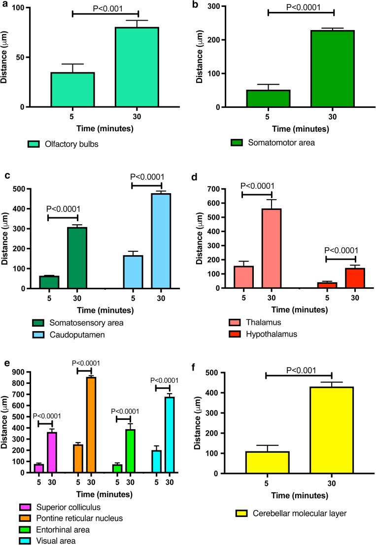
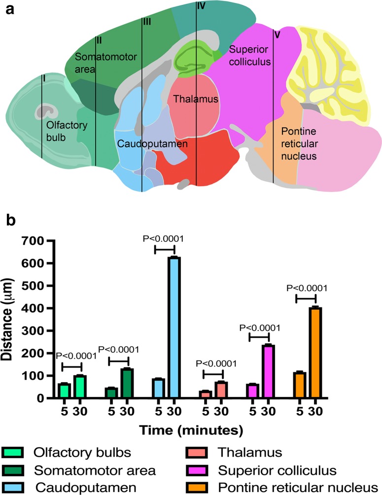
Tracers injected into CSF pass into the brain alongside arteries and out again. This has been recently termed the "glymphatic system" that proposes tracers enter the brain along periarterial "spaces" and leave the brain along the walls of veins. The object of the present study is to test the hypothesis that: (1) tracers from the CSF enter the cerebral cortex along pial-glial basement membranes as there are no perivascular "spaces" around cortical arteries, (2) tracers leave the brain along smooth muscle cell basement membranes that form the Intramural Peri-Arterial Drainage (IPAD) pathways for the elimination of interstitial fluid and solutes from the brain. 2 μL of 100 μM soluble, fluorescent fixable amyloid β (Aβ) were injected into the CSF of the cisterna magna of 6-10 and 24-30 month-old male mice and their brains were examined 5 and 30 min later. At 5 min, immunocytochemistry and confocal microscopy revealed Aβ on the outer aspects of cortical arteries colocalized with α-2 laminin in the pial-glial basement membranes. At 30 min, Aβ was colocalised with collagen IV in smooth muscle cell basement membranes in the walls of cortical arteries corresponding to the IPAD pathways. No evidence for drainage along the walls of veins was found. Measurements of the depth of penetration of tracer were taken from 11 regions of the brain. Maximum depths of penetration of tracer into the brain were achieved in the pons and caudoputamen. Conclusions drawn from the present study are that tracers injected into the CSF enter and leave the brain along separate periarterial basement membrane pathways. The exit route is along IPAD pathways in which Aβ accumulates in cerebral amyloid angiopathy (CAA) in Alzheimer's disease. Results from this study suggest that CSF may be a suitable route for delivery of therapies for neurological diseases, including CAA.
注入 CSF 的示踪剂与动脉一起进入大脑,然后再次流出。这最近被称为“神经胶淋巴系统”,该系统提出示踪剂沿着动脉周围的“空间”进入大脑,并沿着静脉壁离开大脑。本研究的目的是检验以下假设:(1)CSF 中的示踪剂沿着软脑膜-神经胶质基底膜进入大脑皮层,因为在皮质动脉周围没有血管“空间”;(2)示踪剂沿着形成脑内动静脉周围淋巴引流(IPAD)途径的平滑肌细胞基底膜离开大脑,用于从大脑中清除间质液和溶质。将 2 μL 100 μM 可溶性、荧光可固定的淀粉样β(Aβ)注入到 6-10 月龄和 24-30 月龄雄性小鼠的枕大池 CSF 中,然后在 5 分钟和 30 分钟后检查其大脑。在 5 分钟时,免疫细胞化学和共聚焦显微镜显示 Aβ位于皮质动脉外侧面,与软脑膜-神经胶质基底膜中的α-2 层粘连蛋白共定位。在 30 分钟时,Aβ与位于皮质动脉壁平滑肌细胞基底膜中的胶原 IV 共定位,对应于 IPAD 途径。没有发现沿着静脉壁引流的证据。从大脑的 11 个区域测量示踪剂的穿透深度。示踪剂进入大脑的最大深度是在脑桥和尾状核中。从本研究中得出的结论是,注入 CSF 的示踪剂沿着单独的动脉周围基底膜途径进入和离开大脑。出口途径是沿着 IPAD 途径,在阿尔茨海默病的脑淀粉样血管病(CAA)中 Aβ会积聚。本研究的结果表明,CSF 可能是治疗包括 CAA 在内的神经退行性疾病的合适途径。