• 文献检索
  • 文档翻译
  • 深度研究
  • 学术资讯
  • Suppr Zotero 插件Zotero 插件
  • 邀请有礼
  • 套餐&价格
  • 历史记录
应用&插件
Suppr Zotero 插件Zotero 插件浏览器插件Mac 客户端Windows 客户端微信小程序
定价
高级版会员购买积分包购买API积分包
服务
文献检索文档翻译深度研究API 文档MCP 服务
关于我们
关于 Suppr公司介绍联系我们用户协议隐私条款
关注我们

Suppr 超能文献

核心技术专利:CN118964589B侵权必究
粤ICP备2023148730 号-1Suppr @ 2026

文献检索

告别复杂PubMed语法,用中文像聊天一样搜索,搜遍4000万医学文献。AI智能推荐,让科研检索更轻松。

立即免费搜索

文件翻译

保留排版,准确专业,支持PDF/Word/PPT等文件格式,支持 12+语言互译。

免费翻译文档

深度研究

AI帮你快速写综述,25分钟生成高质量综述,智能提取关键信息,辅助科研写作。

立即免费体验

基于 CT 的肺气肿视觉分类:与 COPDGene 研究中死亡率的关联。

CT-based Visual Classification of Emphysema: Association with Mortality in the COPDGene Study.

机构信息

From the Department of Radiology (D.A.L., D.N., T.J., S.M.H.), Division of Biostatistics (C.M.M., C.W., D.C.E.), and Department of Medicine (E.A.R., B.J.M., R.P.B., J.D.C.), National Jewish Health, 1400 Jackson St, Denver, CO 80206; Department of Radiology, Columbia University Medical Center, New York, NY (J.H.M.A.); Department of Diagnostic Radiology, Hôpital Pitié-Salpêtrière, Assistance Publique-Hôpitaux de Paris, Sorbonne Universités, Paris, France (P.A.G.); Department of Diagnostic and Interventional Radiology, University of Heidelberg, Translational Lung Research Center Heidelberg, Heidelberg, Germany (H.U.K.); Division of Pulmonary and Critical Care Medicine, Department of Internal Medicine, University of Michigan Health System, Ann Arbor, Mich (M.K.H., J.L.C.); Department of Epidemiology, Johns Hopkins Bloomberg School of Public Health, Baltimore, Md (T.H.B.); Department of Epidemiology, Colorado School of Public Health, University of Colorado Denver, Anschutz Medical Campus, Aurora, Colo (J.E.H.); Medical Service, VA Ann Arbor Healthcare System, Ann Arbor, Mich (J.L.C.); and Division of Network Medicine, Brigham and Women's Hospital and Harvard Medical School, Boston, Mass (E.K.S.).

出版信息

Radiology. 2018 Sep;288(3):859-866. doi: 10.1148/radiol.2018172294. Epub 2018 May 15.

DOI:10.1148/radiol.2018172294
PMID:29762095
原文链接:https://pmc.ncbi.nlm.nih.gov/articles/PMC6122195/
Abstract

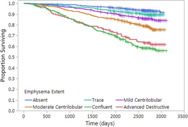
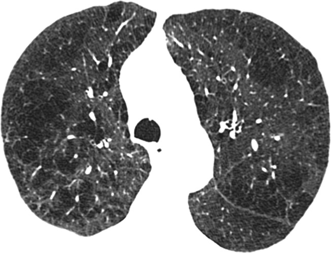
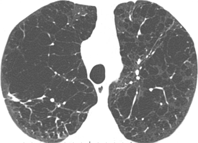
Purpose To determine whether visually assessed patterns of emphysema at CT might provide a simple assessment of mortality risk among cigarette smokers. Materials and Methods Of the first 4000 cigarette smokers consecutively enrolled between 2007 and 2011 in this COPDGene study, 3171 had data available for both visual emphysema CT scores and survival. Each CT scan was retrospectively visually scored by two analysts using the Fleischner Society classification system. Severity of emphysema was also evaluated quantitatively by using percentage lung volume occupied by low-attenuation areas (voxels with attenuation of -950 HU or less) (LAA-950). Median duration of follow-up was 7.4 years. Regression analysis for the relationship between imaging patterns and survival was based on the Cox proportional hazards model, with adjustment for age, race, sex, height, weight, pack-years of cigarette smoking, current smoking status, educational level, LAA-950, and (in a second model) forced expiratory volume in 1 second (FEV). Results Observer agreement in visual scoring was good (weighted κ values, 0.71-0.80). There were 519 deaths in the study cohort. Compared with subjects who did not have visible emphysema, mortality was greater in those with any grade of emphysema beyond trace (adjusted hazard ratios, 1.7, 2.5, 5.0, and 4.1, respectively, for mild centrilobular emphysema, moderate centrilobular emphysema, confluent emphysema, and advanced destructive emphysema, P < .001). This increased mortality generally persisted after adjusting for LAA-950. Conclusion The visual presence and severity of emphysema is associated with significantly increased mortality risk, independent of the quantitative severity of emphysema. Online supplemental material is available for this article.

摘要

目的

确定 CT 上目测肺气肿模式是否可以简单评估吸烟人群的死亡风险。

材料与方法

在这项 COPDGene 研究中,连续纳入了 2007 年至 2011 年间的前 4000 名吸烟者,其中 3171 名吸烟者的 CT 视觉肺气肿评分和生存数据均可用。每个 CT 扫描均由两位分析师使用 Fleischner 学会分类系统进行回顾性视觉评分。使用低衰减区(衰减值低于-950 HU 的体素)(LAA-950)占肺容积的百分比(%)来定量评估肺气肿的严重程度。中位随访时间为 7.4 年。基于 Cox 比例风险模型,对成像模式与生存之间的关系进行回归分析,调整因素包括年龄、种族、性别、身高、体重、吸烟包年数、当前吸烟状态、教育程度、LAA-950 以及(在第二个模型中)1 秒用力呼气量(FEV)。

结果

视觉评分的观察者间一致性良好(加权κ值,0.71-0.80)。研究队列中有 519 例死亡。与没有可见肺气肿的患者相比,轻度小叶中心型肺气肿、中度小叶中心型肺气肿、融合型肺气肿和晚期破坏性肺气肿患者的死亡率分别增加了 1.7 倍、2.5 倍、5.0 倍和 4.1 倍(调整后的危险比,分别为 1.7、2.5、5.0 和 4.1,P <.001)。调整 LAA-950 后,这种死亡率的增加仍普遍存在。

结论

肺气肿的视觉存在和严重程度与显著增加的死亡风险相关,独立于肺气肿的定量严重程度。本文提供了在线补充材料。

https://cdn.ncbi.nlm.nih.gov/pmc/blobs/9366/6122195/86e476c4414a/radiol.2018172294.fig2.jpg
https://cdn.ncbi.nlm.nih.gov/pmc/blobs/9366/6122195/5f40c0683fb0/radiol.2018172294.fig1a.jpg
https://cdn.ncbi.nlm.nih.gov/pmc/blobs/9366/6122195/34b72fab06f3/radiol.2018172294.fig1b.jpg
https://cdn.ncbi.nlm.nih.gov/pmc/blobs/9366/6122195/1fb28ade8a1f/radiol.2018172294.fig1c.jpg
https://cdn.ncbi.nlm.nih.gov/pmc/blobs/9366/6122195/31026db7fd31/radiol.2018172294.fig1d.jpg
https://cdn.ncbi.nlm.nih.gov/pmc/blobs/9366/6122195/d692b4387982/radiol.2018172294.fig1e.jpg
https://cdn.ncbi.nlm.nih.gov/pmc/blobs/9366/6122195/d0f4d20b5021/radiol.2018172294.fig1f.jpg
https://cdn.ncbi.nlm.nih.gov/pmc/blobs/9366/6122195/86e476c4414a/radiol.2018172294.fig2.jpg

相似文献

1
CT-based Visual Classification of Emphysema: Association with Mortality in the COPDGene Study.基于 CT 的肺气肿视觉分类:与 COPDGene 研究中死亡率的关联。
Radiology. 2018 Sep;288(3):859-866. doi: 10.1148/radiol.2018172294. Epub 2018 May 15.
2
Fleischner Society Visual Emphysema CT Patterns Help Predict Progression of Emphysema in Current and Former Smokers: Results from the COPDGene Study.弗莱舍纳社会视觉肺气肿 CT 模式有助于预测当前和曾经吸烟者肺气肿的进展:COPDGene 研究结果。
Radiology. 2021 Feb;298(2):441-449. doi: 10.1148/radiol.2020200563. Epub 2020 Dec 15.
3
Emphysema Progression at CT by Deep Learning Predicts Functional Impairment and Mortality: Results from the COPDGene Study.深度学习预测 CT 肺气肿进展与功能障碍和死亡率的关系:COPDGene 研究结果。
Radiology. 2022 Sep;304(3):672-679. doi: 10.1148/radiol.213054. Epub 2022 May 17.
4
Visual Emphysema at Chest CT in GOLD Stage 0 Cigarette Smokers Predicts Disease Progression: Results from the COPDGene Study.胸部 CT 肺气肿在 GOLD 分期 0 期的吸烟人群中预测疾病进展:COPDGene 研究结果。
Radiology. 2020 Sep;296(3):641-649. doi: 10.1148/radiol.2020192429. Epub 2020 Jul 7.
5
Quantitative computed tomography measurements to evaluate airway disease in chronic obstructive pulmonary disease: Relationship to physiological measurements, clinical index and visual assessment of airway disease.定量计算机断层扫描测量评估慢性阻塞性肺疾病中的气道疾病:与气道疾病的生理测量、临床指标及视觉评估的关系
Eur J Radiol. 2016 Nov;85(11):2144-2151. doi: 10.1016/j.ejrad.2016.09.010. Epub 2016 Sep 13.
6
Deep Learning Enables Automatic Classification of Emphysema Pattern at CT.深度学习可实现 CT 肺气肿模式的自动分类。
Radiology. 2020 Feb;294(2):434-444. doi: 10.1148/radiol.2019191022. Epub 2019 Dec 3.
7
Spatial Dependence of CT Emphysema in Chronic Obstructive Pulmonary Disease Quantified by Using Join-Count Statistics.利用连接计数统计量定量分析慢性阻塞性肺疾病 CT 肺气肿的空间依赖性。
Radiology. 2021 Dec;301(3):702-709. doi: 10.1148/radiol.2021210198. Epub 2021 Sep 14.
8
Relationships between diffusing capacity for carbon monoxide (DLCO), and quantitative computed tomography measurements and visual assessment for chronic obstructive pulmonary disease.一氧化碳弥散量(DLCO)与慢性阻塞性肺疾病的定量计算机断层扫描测量及视觉评估之间的关系。
Eur J Radiol. 2015 May;84(5):980-5. doi: 10.1016/j.ejrad.2015.01.010. Epub 2015 Jan 22.
9
Relationship between Emphysema Progression at CT and Mortality in Ever-Smokers: Results from the COPDGene and ECLIPSE Cohorts.CT 肺气肿进展与持续吸烟者死亡率的关系:COPDGene 和 ECLIPSE 队列研究结果。
Radiology. 2021 Apr;299(1):222-231. doi: 10.1148/radiol.2021203531. Epub 2021 Feb 16.
10
A combined pulmonary-radiology workshop for visual evaluation of COPD: study design, chest CT findings and concordance with quantitative evaluation.COPD 的肺放射联合视觉评估研讨会:研究设计、胸部 CT 结果及与定量评估的一致性。
COPD. 2012 Apr;9(2):151-9. doi: 10.3109/15412555.2012.654923. Epub 2012 Mar 19.

引用本文的文献

1
Emphysema progression risk in COPD using a localized foundational model of density evolution.使用密度演变的局部基础模型评估慢性阻塞性肺疾病(COPD)中肺气肿进展风险
NPJ Digit Med. 2025 Aug 28;8(1):556. doi: 10.1038/s41746-025-01917-3.
2
Differentiating Emphysema From Emphysema-Dominated COPD Patients with CT Imaging Feature and Machine Learning.利用CT成像特征和机器学习鉴别肺气肿与以肺气肿为主的慢性阻塞性肺疾病患者
Int J Chron Obstruct Pulmon Dis. 2025 Jul 25;20:2615-2628. doi: 10.2147/COPD.S527914. eCollection 2025.
3
Towards a more realistic anthropomorphic chest phantom using 3D-printed and cork-integrated components.

本文引用的文献

1
Visual Assessment of Chest Computed Tomographic Images Is Independently Useful for Genetic Association Analysis in Studies of Chronic Obstructive Pulmonary Disease.胸部计算机断层扫描图像的视觉评估可独立用于慢性阻塞性肺疾病研究中的遗传关联分析。
Ann Am Thorac Soc. 2017 Jan;14(1):33-40. doi: 10.1513/AnnalsATS.201606-427OC.
2
Per cent emphysema is associated with respiratory and lung cancer mortality in the general population: a cohort study.肺气肿百分比与普通人群的呼吸和肺癌死亡率相关:一项队列研究。
Thorax. 2016 Jul;71(7):624-32. doi: 10.1136/thoraxjnl-2015-207822. Epub 2016 Apr 5.
3
Clinical and Radiologic Disease in Smokers With Normal Spirometry.
利用3D打印和软木集成部件制作更逼真的拟人化胸部体模。
Med Phys. 2025 Jul;52(7):e17956. doi: 10.1002/mp.17956.
4
Radiological Changes in Chest Computed Tomography Findings of School Cooks: A Comparative Study With Age-matched Controls.学校厨师胸部计算机断层扫描结果的影像学变化:与年龄匹配对照组的比较研究
Saf Health Work. 2025 Jun;16(2):228-235. doi: 10.1016/j.shaw.2025.01.006. Epub 2025 Jan 30.
5
Respiratory symptoms associated with a new lobe-based bronchial scoring system in an urban Chinese low-dose CT screening population.中国城市低剂量CT筛查人群中与一种基于肺叶的新支气管评分系统相关的呼吸道症状
Eur Radiol. 2025 Jun 13. doi: 10.1007/s00330-025-11712-z.
6
Analysis of factors influencing hookwire dislodgement in CT-guided hookwire localization: a retrospective study using variable importance analysis with a random forest model.CT引导下导丝定位中影响导丝移位因素的分析:一项使用随机森林模型进行变量重要性分析的回顾性研究
PeerJ. 2025 Apr 16;13:e19231. doi: 10.7717/peerj.19231. eCollection 2025.
7
The association and impact of radiographic, pathological emphysema and spirometric airway obstruction on patients with resectable lung adenocarcinoma.影像学、病理学肺气肿及肺功能测定气道阻塞对可切除肺腺癌患者的关联及影响
Respir Res. 2025 Apr 16;26(1):151. doi: 10.1186/s12931-025-03225-6.
8
The Use of CT Densitometry for the Assessment of Emphysema in Clinical Trials: A Position Paper from the Fleischner Society.CT密度测定法在临床试验中用于评估肺气肿:来自弗莱施纳学会的立场文件。
Am J Respir Crit Care Med. 2025 Mar 24;211(5):709-28. doi: 10.1164/rccm.202410-2012SO.
9
Non-cancerous CT findings as predictors of survival outcome in advanced non-small cell lung cancer patients treated with first-generation EGFR-TKIs.非癌性CT表现作为接受第一代表皮生长因子受体酪氨酸激酶抑制剂(EGFR-TKIs)治疗的晚期非小细胞肺癌患者生存结果的预测指标
PLoS One. 2025 Feb 5;20(2):e0313577. doi: 10.1371/journal.pone.0313577. eCollection 2025.
10
Low-Dose Computed Tomography Scan Features Are Associated With Annual Risk of Hospitalization.低剂量计算机断层扫描特征与年度住院风险相关。
Ann Thorac Surg Short Rep. 2023 Jul 15;1(4):558-561. doi: 10.1016/j.atssr.2023.06.011. eCollection 2023 Dec.
肺功能正常的吸烟者的临床和放射学疾病
JAMA Intern Med. 2015 Sep;175(9):1539-49. doi: 10.1001/jamainternmed.2015.2735.
4
CT-Definable Subtypes of Chronic Obstructive Pulmonary Disease: A Statement of the Fleischner Society.慢性阻塞性肺疾病的CT可定义亚型:弗莱施纳学会声明
Radiology. 2015 Oct;277(1):192-205. doi: 10.1148/radiol.2015141579. Epub 2015 May 11.
5
Paraseptal emphysema: Prevalence and distribution on CT and association with interstitial lung abnormalities.间隔旁型肺气肿:CT上的患病率、分布及其与间质性肺异常的关联
Eur J Radiol. 2015 Jul;84(7):1413-8. doi: 10.1016/j.ejrad.2015.03.010. Epub 2015 Mar 18.
6
Visual Assessment of CT Findings in Smokers With Nonobstructed Spirometric Abnormalities in The COPDGene Study.COPDGene研究中肺功能正常的吸烟者CT表现的视觉评估
Chronic Obstr Pulm Dis. 2014;1(1):88-96. doi: 10.15326/jcopdf.1.1.2013.0001#sthash.L0atdpjM.dpuf.
7
Epidemiology, genetics, and subtyping of preserved ratio impaired spirometry (PRISm) in COPDGene.慢性阻塞性肺疾病基因研究(COPDGene)中肺量计保存率受损(PRISm)的流行病学、遗传学及亚型分析
Respir Res. 2014 Aug 6;15(1):89. doi: 10.1186/s12931-014-0089-y.
8
Cardiovascular disease is associated with COPD severity and reduced functional status and quality of life.心血管疾病与慢性阻塞性肺疾病的严重程度、功能状态降低及生活质量下降相关。
COPD. 2014 Sep;11(5):546-51. doi: 10.3109/15412555.2014.898029. Epub 2014 May 15.
9
Pulmonary emphysema subtypes on computed tomography: the MESA COPD study.计算机断层扫描显示的肺气肿亚型:MESA COPD 研究。
Am J Med. 2014 Jan;127(1):94.e7-23. doi: 10.1016/j.amjmed.2013.09.020. Epub 2013 Oct 9.
10
Distinct quantitative computed tomography emphysema patterns are associated with physiology and function in smokers.不同定量 CT 肺气肿模式与吸烟者的生理学和功能有关。
Am J Respir Crit Care Med. 2013 Nov 1;188(9):1083-90. doi: 10.1164/rccm.201305-0873OC.