Department Biology I, Ultrastructural Research, , Ludwig-Maximilians-University Munich, 82152, Planegg-Martinsried, Germany.
Histochem Cell Biol. 2018 Aug;150(2):149-170. doi: 10.1007/s00418-018-1681-x. Epub 2018 May 23.
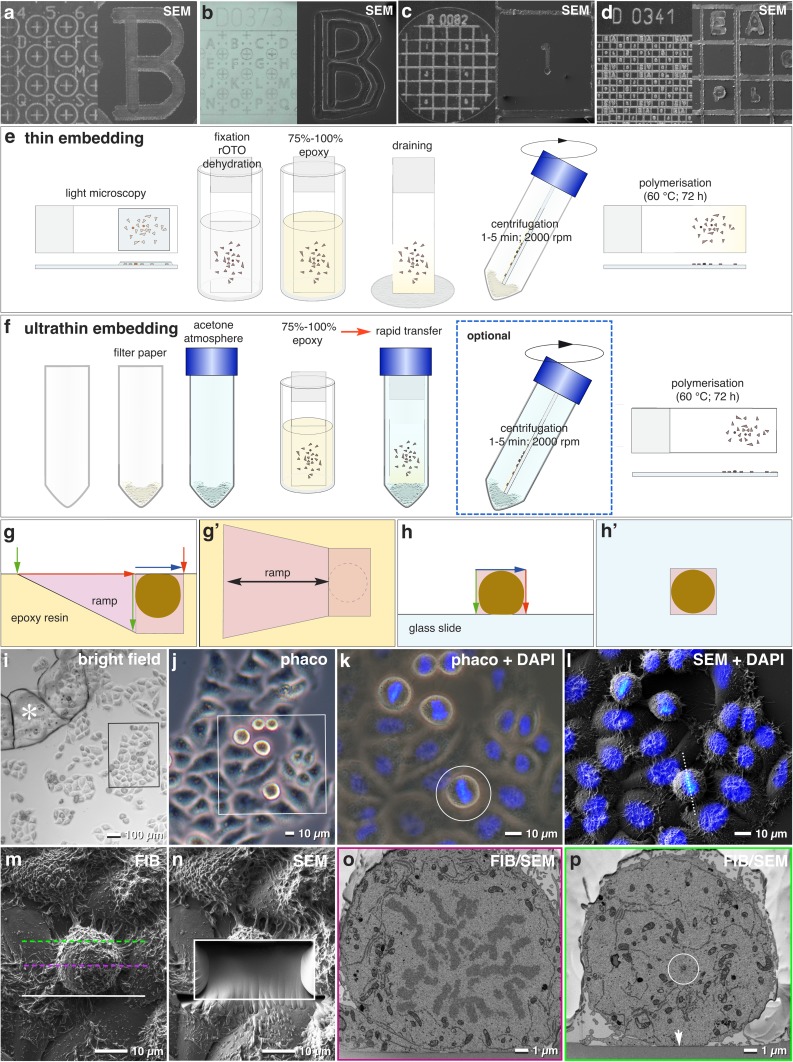
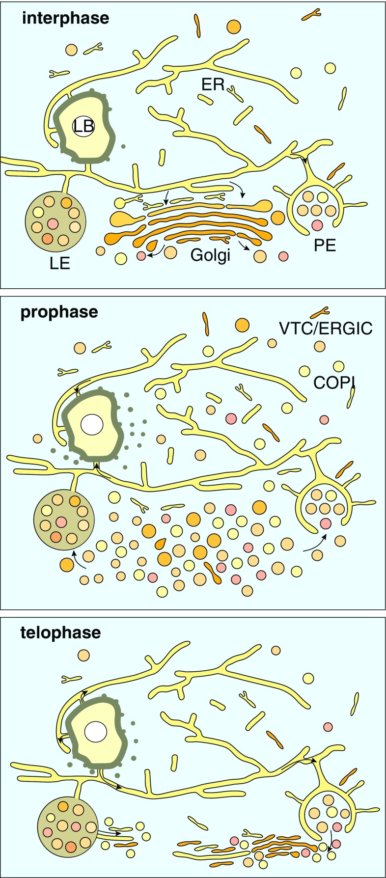
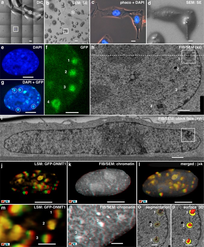
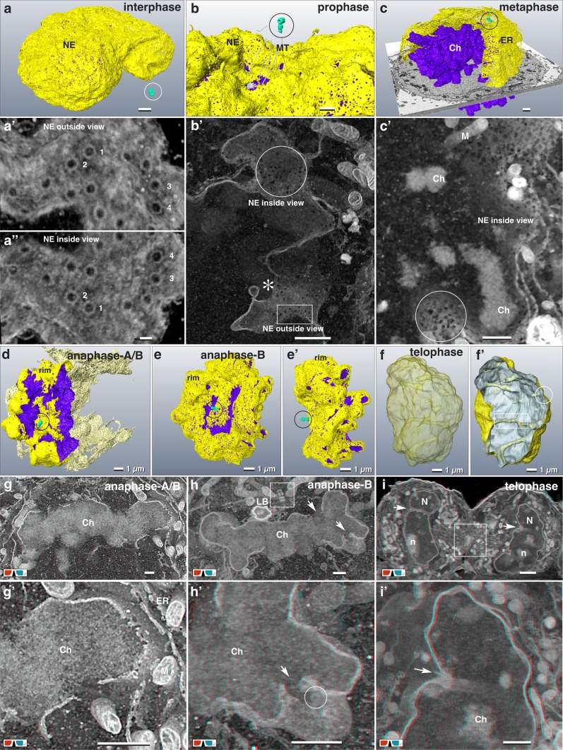
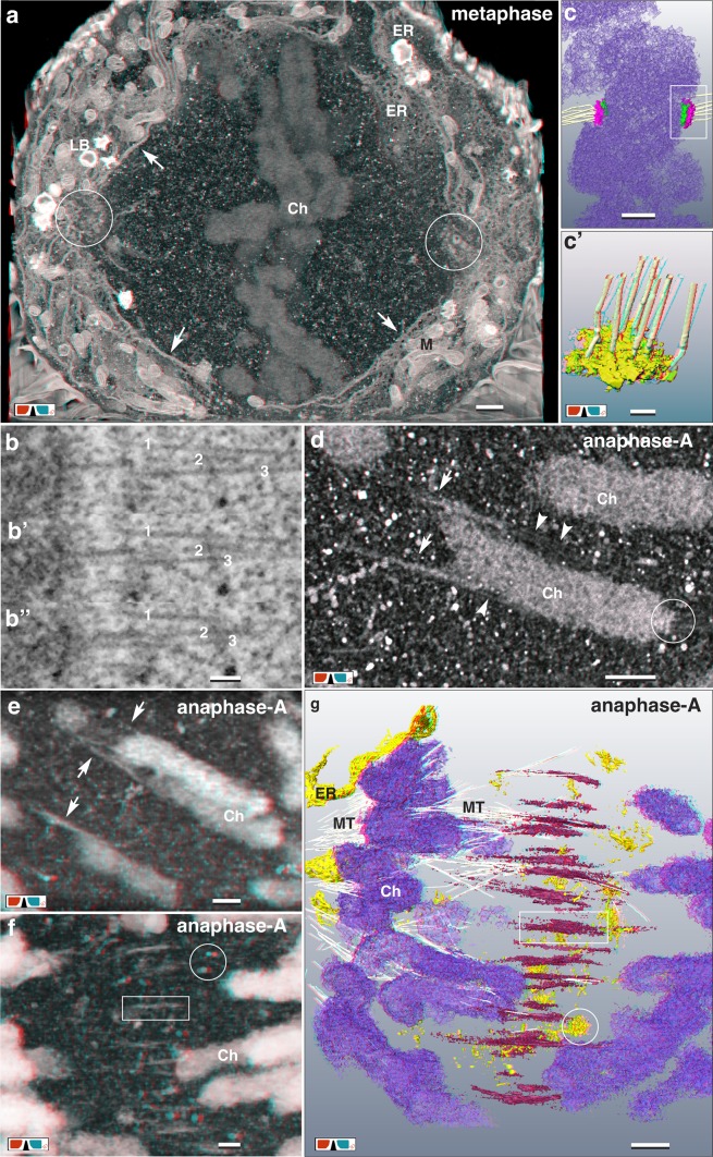
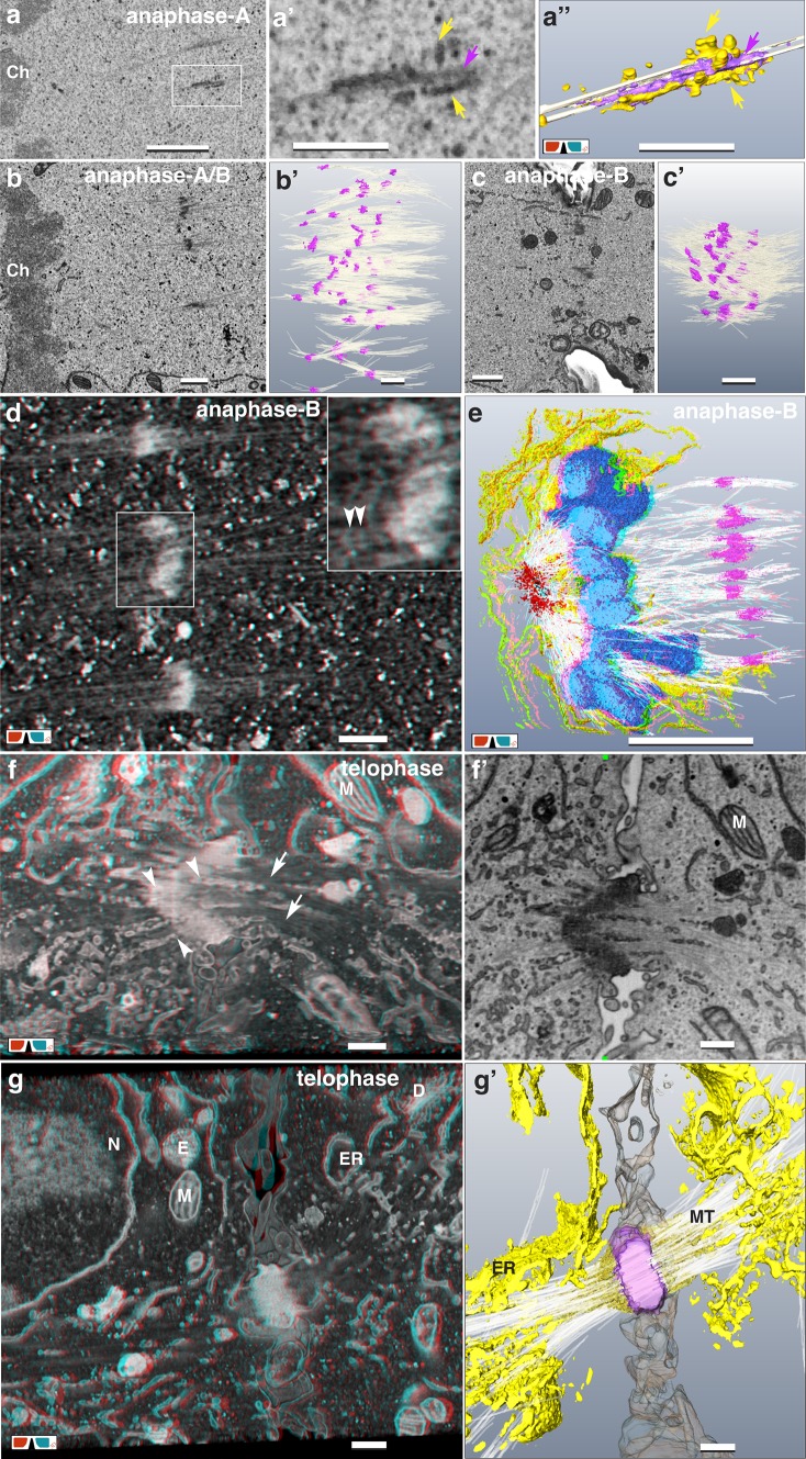
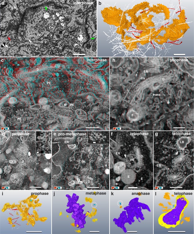
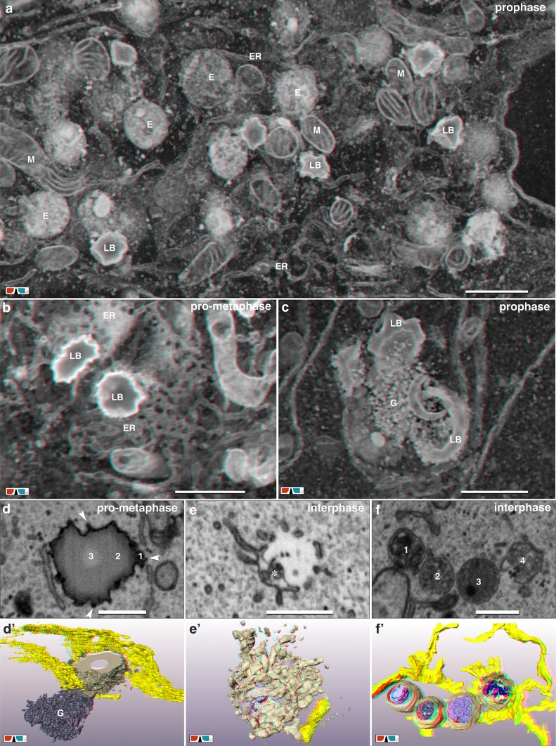
A portfolio is presented documenting economic, high-resolution correlative focused ion beam scanning electron microscopy (FIB/SEM) in routine, comprising: (i) the use of custom-labeled slides and coverslips, (ii) embedding of cells in thin, or ultra-thin resin layers for correlative light and electron microscopy (CLEM) and (iii) the claim to reach the highest resolution possible with FIB/SEM in xyz. Regions of interest (ROIs) defined in light microscope (LM), can be relocated quickly and precisely in SEM. As proof of principle, HeLa cells were investigated in 3D context at all stages of the cell cycle, documenting ultrastructural changes during mitosis: nuclear envelope breakdown and reassembly, Golgi degradation and reconstitution and the formation of the midzone and midbody.
现提供一份文件,记录经济实惠、高分辨率的相关聚焦离子束扫描电子显微镜(FIB/SEM)在常规应用中的情况,包括:(i)使用定制标记的载玻片和盖玻片,(ii)将细胞嵌入薄或超薄树脂层中,用于相关的光和电子显微镜(CLEM),以及(iii)声称在 xyz 方向上达到 FIB/SEM 的最高分辨率。在光显微镜(LM)中定义的感兴趣区域(ROI)可以在 SEM 中快速且精确地重新定位。作为原理验证,在细胞周期的所有阶段对 HeLa 细胞进行了 3D 研究,记录了有丝分裂过程中的超微结构变化:核膜的崩解和重组、高尔基体的降解和重建以及中体和中体的形成。