• 文献检索
  • 文档翻译
  • 深度研究
  • 学术资讯
  • Suppr Zotero 插件Zotero 插件
  • 邀请有礼
  • 套餐&价格
  • 历史记录
应用&插件
Suppr Zotero 插件Zotero 插件浏览器插件Mac 客户端Windows 客户端微信小程序
定价
高级版会员购买积分包购买API积分包
服务
文献检索文档翻译深度研究API 文档MCP 服务
关于我们
关于 Suppr公司介绍联系我们用户协议隐私条款
关注我们

Suppr 超能文献

核心技术专利:CN118964589B侵权必究
粤ICP备2023148730 号-1Suppr @ 2026

文献检索

告别复杂PubMed语法,用中文像聊天一样搜索,搜遍4000万医学文献。AI智能推荐,让科研检索更轻松。

立即免费搜索

文件翻译

保留排版,准确专业,支持PDF/Word/PPT等文件格式,支持 12+语言互译。

免费翻译文档

深度研究

AI帮你快速写综述,25分钟生成高质量综述,智能提取关键信息,辅助科研写作。

立即免费体验

通过增强扫描电子显微镜对精子超微结构的新见解

New Insights Into Sperm Ultrastructure Through Enhanced Scanning Electron Microscopy.

作者信息

Korneev Denis, Merriner D Jo, Gervinskas Gediminas, de Marco Alex, O'Bryan Moira K

机构信息

School of Biological Sciences, Monash University, Melbourne, VIC, Australia.

Department of Biochemistry and Molecular Biology, Biomedicine Discovery Institute, Monash University, Melbourne, VIC, Australia.

出版信息

Front Cell Dev Biol. 2021 Apr 22;9:672592. doi: 10.3389/fcell.2021.672592. eCollection 2021.

DOI:10.3389/fcell.2021.672592
PMID:33968944
原文链接:https://pmc.ncbi.nlm.nih.gov/articles/PMC8100687/
Abstract

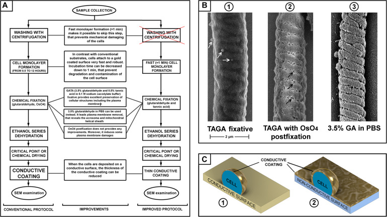
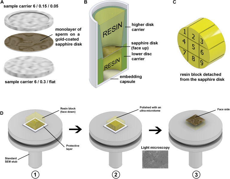
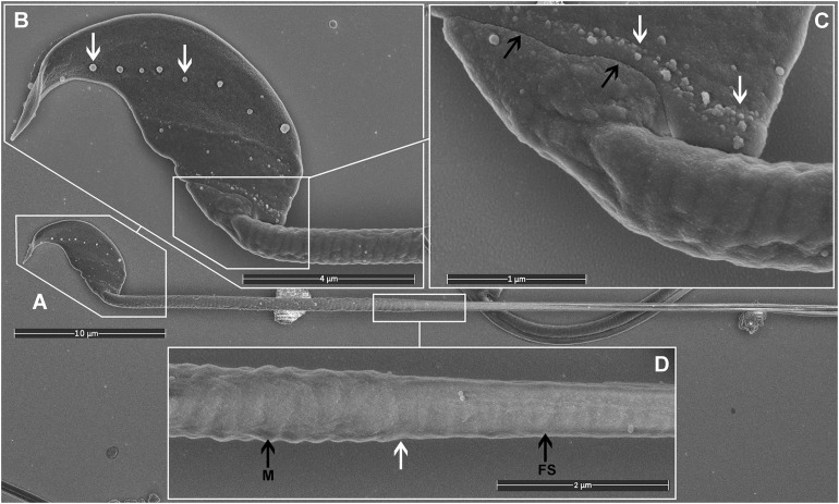
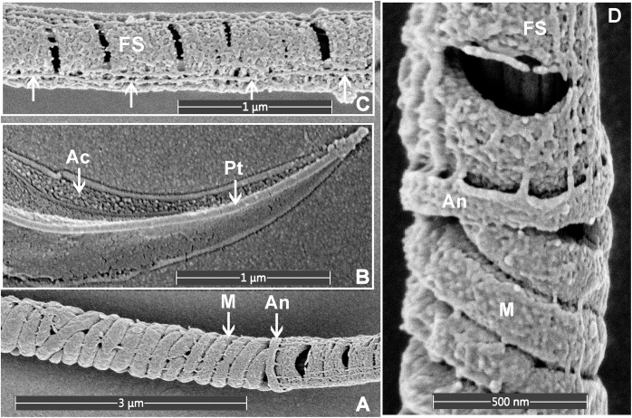
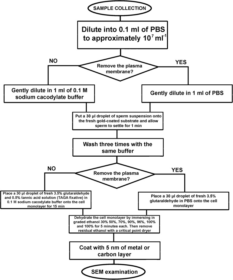
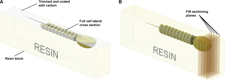
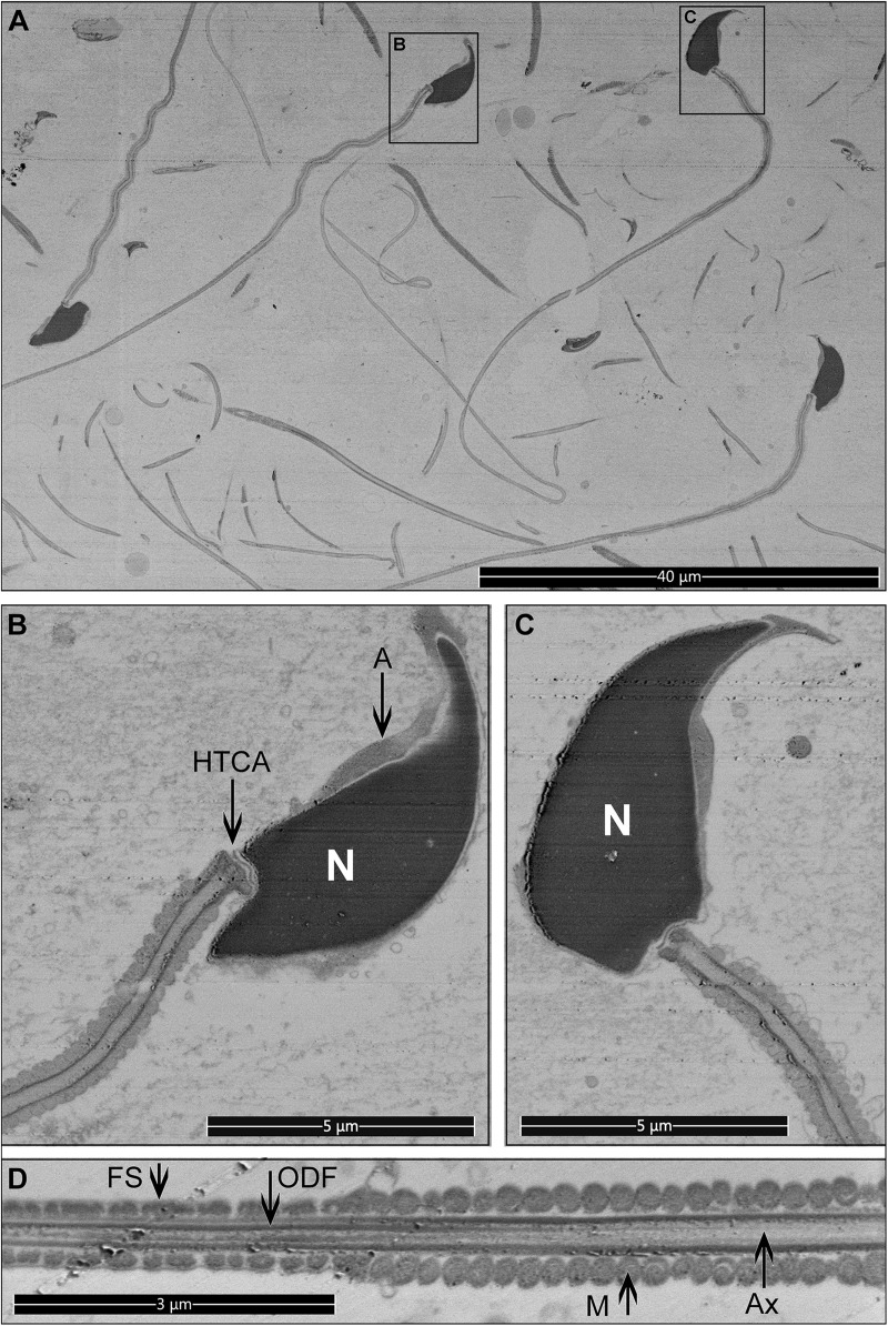
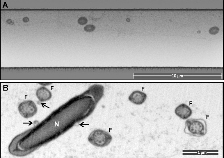
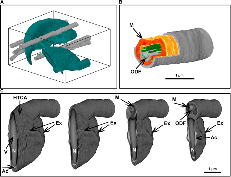
The analysis of spermatozoa morphology is fundamental to understand male fertility and the etiology of infertility. Traditionally scanning electron microscopy (SEM) has been used to define surface topology. Recently, however, it has become a critical tool for three-dimensional analysis of internal cellular ultrastructure. Modern SEM provides nanometer-scale resolution, but the meaningfulness of such information is proportional to the quality of the sample preservation. In this study, we demonstrate that sperm quickly and robustly adhere to gold-coated surfaces. Leveraging this property, we developed three step-by-step protocols fulfilling different needs for sperm imaging: chemically fixed monolayers for SEM examination of the external morphology, and two high-pressure freezing-based protocols for fast SEM examination of full cell internal morphology and focused ion-beam SEM tomography. These analyses allow previously unappreciated insights into mouse sperm ultrastructure, including the identification of novel structures within the fibrous sheath and domain-specific interactions between the plasma membrane and exosome-like structures.

摘要

精子形态分析对于理解男性生育能力和不育症病因至关重要。传统上,扫描电子显微镜(SEM)一直用于定义表面拓扑结构。然而,最近它已成为细胞内部超微结构三维分析的关键工具。现代SEM提供纳米级分辨率,但此类信息的意义与样品保存质量成正比。在本研究中,我们证明精子能快速且牢固地附着在镀金表面。利用这一特性,我们开发了三个逐步方案,以满足精子成像的不同需求:用于外部形态SEM检查的化学固定单层,以及用于全细胞内部形态快速SEM检查和聚焦离子束SEM断层扫描的两个基于高压冷冻的方案。这些分析使我们对小鼠精子超微结构有了前所未有的深入了解,包括在纤维鞘内识别新结构以及质膜与外泌体样结构之间的区域特异性相互作用。

https://cdn.ncbi.nlm.nih.gov/pmc/blobs/f260/8100687/d2aa1986812d/fcell-09-672592-g009.jpg
https://cdn.ncbi.nlm.nih.gov/pmc/blobs/f260/8100687/a6e47e0eff1c/fcell-09-672592-g001.jpg
https://cdn.ncbi.nlm.nih.gov/pmc/blobs/f260/8100687/6dea5516adca/fcell-09-672592-g002.jpg
https://cdn.ncbi.nlm.nih.gov/pmc/blobs/f260/8100687/aaa63e3e3a59/fcell-09-672592-g003.jpg
https://cdn.ncbi.nlm.nih.gov/pmc/blobs/f260/8100687/52d4cfd76265/fcell-09-672592-g004.jpg
https://cdn.ncbi.nlm.nih.gov/pmc/blobs/f260/8100687/01f39e5dc6fe/fcell-09-672592-g005.jpg
https://cdn.ncbi.nlm.nih.gov/pmc/blobs/f260/8100687/02a684818695/fcell-09-672592-g006.jpg
https://cdn.ncbi.nlm.nih.gov/pmc/blobs/f260/8100687/2bee2a28a9eb/fcell-09-672592-g007.jpg
https://cdn.ncbi.nlm.nih.gov/pmc/blobs/f260/8100687/3f7ddc158d32/fcell-09-672592-g008.jpg
https://cdn.ncbi.nlm.nih.gov/pmc/blobs/f260/8100687/d2aa1986812d/fcell-09-672592-g009.jpg

相似文献

1
New Insights Into Sperm Ultrastructure Through Enhanced Scanning Electron Microscopy.通过增强扫描电子显微镜对精子超微结构的新见解
Front Cell Dev Biol. 2021 Apr 22;9:672592. doi: 10.3389/fcell.2021.672592. eCollection 2021.
2
FIB-SEM imaging properties of Drosophila melanogaster tissues embedded in Lowicryl HM20.在低聚甲醛 HM20 中包埋的果蝇组织的 FIB-SEM 成像特性。
J Microsc. 2019 Feb;273(2):91-104. doi: 10.1111/jmi.12764. Epub 2018 Nov 12.
3
Ultrastructural description of fresh and frozen/thawed sperm derived from collared peccaries (Pecari tajacu Linnaeus, 1,758).白唇野猪(Pecari tajacu Linnaeus,1758年)新鲜及冷冻/解冻精子的超微结构描述。
Microsc Res Tech. 2018 Nov;81(11):1301-1309. doi: 10.1002/jemt.23138. Epub 2018 Oct 8.
4
Comparative analysis of mammalian sperm ultrastructure reveals relationships between sperm morphology, mitochondrial functions and motility.哺乳动物精子超微结构的比较分析揭示了精子形态、线粒体功能和运动能力之间的关系。
Reprod Biol Endocrinol. 2019 Aug 15;17(1):66. doi: 10.1186/s12958-019-0510-y.
5
New advances in scanning microscopy and its application to study parasitic protozoa.扫描显微镜的新进展及其在研究寄生原生动物中的应用。
Exp Parasitol. 2018 Jul;190:10-33. doi: 10.1016/j.exppara.2018.04.018. Epub 2018 Apr 25.
6
Three-Dimensional Ultrastructural Analysis of the Head-Most Mitochondrial Roots of Mice Spermatozoa Using Focused Ion Beam/Scanning Electron Microscopy (FIB/SEM) Tomography.使用聚焦离子束/扫描电子显微镜(FIB/SEM)断层扫描技术对小鼠精子头部线粒体根的三维超微结构分析。
Microsc Microanal. 2023 Jul 25;29(4):1460-1466. doi: 10.1093/micmic/ozad055.
7
High-Resolution 3D Imaging of Megakaryocytes Using Focused Ion Beam-Scanning Electron Microscopy.使用聚焦离子束扫描电子显微镜对巨核细胞进行高分辨率三维成像
Methods Mol Biol. 2018;1812:217-231. doi: 10.1007/978-1-4939-8585-2_13.
8
The importance of transmission electron microscopy analysis of spermatozoa: Diagnostic applications and basic research.精子透射电子显微镜分析的重要性:诊断应用与基础研究
Syst Biol Reprod Med. 2016 Jun;62(3):171-83. doi: 10.3109/19396368.2016.1155242. Epub 2016 Mar 16.
9
Micromorphological and ultrastructural description of spermatozoa from squirrel monkeys ( Osgood, 1916).来自松鼠猴(Osgood,1916)精子的微观形态和超微结构描述。
Zygote. 2020 Jun;28(3):203-207. doi: 10.1017/S0967199419000868. Epub 2020 Jan 14.
10
Preparation of biological monolayers for producing high-resolution scanning electron micrographs.生物单层的制备用于产生高分辨率扫描电子显微镜照片。
PLoS One. 2022 Jul 8;17(7):e0266943. doi: 10.1371/journal.pone.0266943. eCollection 2022.

引用本文的文献

1
A novel mechanism of sperm midpiece epididymal maturation and the role of CCDC112 in sperm midpiece formation and establishing an optimal flagella waveform.精子中段附睾成熟的一种新机制以及CCDC112在精子中段形成和建立最佳鞭毛波形中的作用。
Cell Commun Signal. 2025 Jul 1;23(1):319. doi: 10.1186/s12964-025-02320-x.
2
Critical role of Spatio-Temporally Regulated Maternal RNAs in Zebrafish Embryogenesis.时空调控的母源RNA在斑马鱼胚胎发育中的关键作用
bioRxiv. 2024 Nov 8:2024.11.07.622483. doi: 10.1101/2024.11.07.622483.
3
Impact of air pollution and heavy metal exposure on sperm quality: A clinical prospective research study.

本文引用的文献

1
The Role of the Epididymis and the Contribution of Epididymosomes to Mammalian Reproduction.附睾的作用和附睾小体对哺乳动物生殖的贡献。
Int J Mol Sci. 2020 Jul 29;21(15):5377. doi: 10.3390/ijms21155377.
2
Sperm competition and the evolution of sperm form and function in mammals.精子竞争与哺乳动物精子形态和功能的进化
Reprod Domest Anim. 2019 Oct;54 Suppl 4:14-21. doi: 10.1111/rda.13552.
3
The contribution of epididymosomes to the sperm small RNA profile.附睾小体对精子小 RNA 谱的贡献。
空气污染和重金属暴露对精子质量的影响:一项临床前瞻性研究。
Toxicol Rep. 2024 Aug 8;13:101708. doi: 10.1016/j.toxrep.2024.101708. eCollection 2024 Dec.
4
AXDND1 is required to balance spermatogonial commitment and for sperm tail formation in mice and humans.AXDND1 对于维持精原细胞的定向分化以及精子尾部的形成在小鼠和人类中都发挥着重要作用。
Cell Death Dis. 2024 Jul 12;15(7):499. doi: 10.1038/s41419-024-06874-5.
5
Genetic mutation of results in male infertility due to abnormal sperm tail composition.基因突变导致精子尾部组成异常,从而引起男性不育。
Life Sci Alliance. 2024 Apr 3;7(6). doi: 10.26508/lsa.202302452. Print 2024 Jun.
6
The Cby3/ciBAR1 complex positions the annulus along the sperm flagellum during spermiogenesis.Cby3/ciBAR1 复合物在精子发生过程中使环沿着精子鞭毛定位。
J Cell Biol. 2024 Mar 4;223(3). doi: 10.1083/jcb.202307147. Epub 2024 Jan 10.
Reproduction. 2019 Jun;157(6):R209-R223. doi: 10.1530/REP-18-0480.
4
Precise and economic FIB/SEM for CLEM: with 2 nm voxels through mitosis.精准且经济的 FIB/SEM 用于 CLEM:通过有丝分裂实现 2nm 体素。
Histochem Cell Biol. 2018 Aug;150(2):149-170. doi: 10.1007/s00418-018-1681-x. Epub 2018 May 23.
5
Preparation and observation methods can produce misleading artefacts in human sperm ultrastructural morphology.制备和观察方法可能会在人类精子超微结构形态中产生误导性假象。
Andrologia. 2018 Sep;50(7):e13043. doi: 10.1111/and.13043. Epub 2018 May 7.
6
Proteomic analysis of good- and poor-quality human sperm demonstrates that several proteins are routinely aberrantly regulated.蛋白质组学分析优质和劣质人精子表明,有几种蛋白质经常出现异常调节。
Biol Reprod. 2018 Aug 1;99(2):395-408. doi: 10.1093/biolre/iox166.
7
CatSperζ regulates the structural continuity of sperm Ca signaling domains and is required for normal fertility.精子阳离子通道蛋白ζ调节精子钙信号域的结构连续性,是正常生育所必需的。
Elife. 2017 Feb 23;6:e23082. doi: 10.7554/eLife.23082.
8
Boar seminal plasma exosomes maintain sperm function by infiltrating into the sperm membrane.公猪精浆外泌体通过渗入精子膜来维持精子功能。
Oncotarget. 2016 Sep 13;7(37):58832-58847. doi: 10.18632/oncotarget.11315.
9
Ultrastructural Morphology of Sperm from Human Globozoospermia.人类圆头精子症患者精子的超微结构形态
Biomed Res Int. 2015;2015:798754. doi: 10.1155/2015/798754. Epub 2015 Sep 8.
10
Phosphopeptide analysis of rodent epididymal spermatozoa.啮齿动物附睾精子的磷酸肽分析。
J Vis Exp. 2014 Dec 30(94):51546. doi: 10.3791/51546.